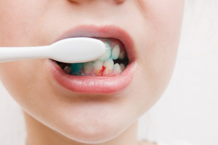
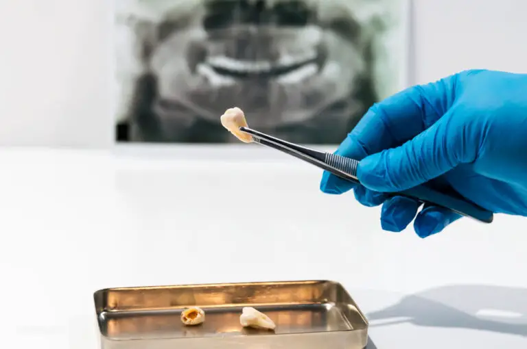
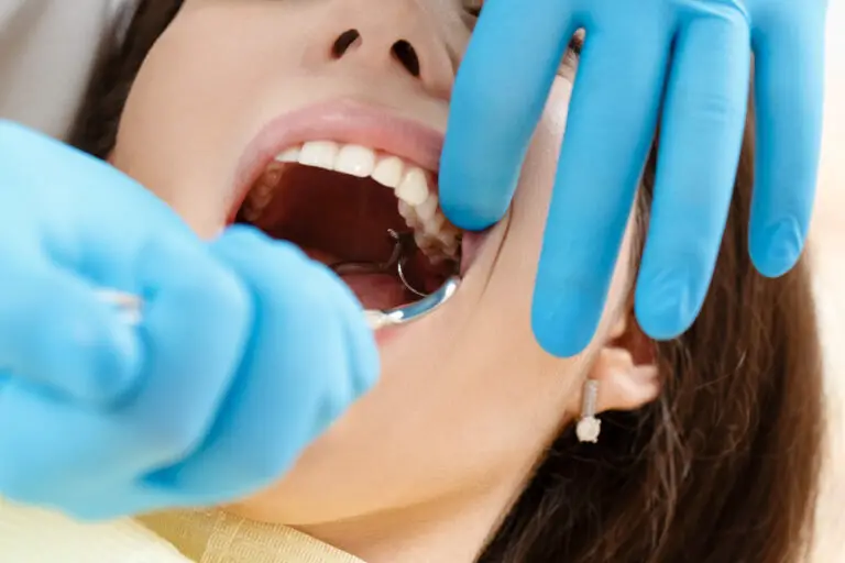
Inside each tooth is a living pulp organ that requires healthy blood flow to survive. The blood vessels extending through the root and pulp chamber supply oxygen, nutrients and infection-fighting cells that keep the tooth alive. When trauma, decay or disease disrupts this circulation, the tooth is at high risk for infection, pain and eventual loss. But some emerging dental treatments aim to revive and restore blood supply to compromised teeth. Regenerating tiny blood vessels or engineering new pulp tissue could potentially rescue damaged teeth. However, many challenges remain before tooth revascularization therapies become clinically practical.
Detailed dental anatomy and physiology

Teeth consist of multiple complex living tissues supported by a nourishing blood supply. Here is a more in-depth look at the anatomy relevant to blood flow:
- Pulp – The pulp contains arteries, veins, lymph vessels, nerves and connective tissues. It extends from the pulp chamber in the crown down through root canals to the root apex.
- Dentin – Dentin makes up the bulk of the tooth surrounding the pulp. It contains thousands of tiny fluid-filled tubules extending from the pulp to the enamel.
- Cementum – Cementum covers the dentin root surface. It provides attachment for the periodontal ligament fibers.
- Periodontal ligament – This connective tissue surrounds and supports the tooth root while also providing avenues for nerve fibers and tiny blood vessels that nourish the cementum.
- Alveolar bone – The socket walls contain spongy bone with marrow spaces, nerves and a network of blood vessels. These supply the PDL and cementum.
- Gingiva – The gums contain the gingival plexus of capillaries nourished by branches of the maxillary and mandibular arteries. Tiny vessels extend into the PDL.
- Nerves – Nerve fibers branch from the trigeminal nerve to reach the pulp and dentin tubules, cementum, PDL and gum tissues. They provide tooth sensitivity.
- Lymphatic drainage – Lymphatic vessels parallel blood circulation to drain fluid and immune cells from the pulp, PDL and oral mucosal tissues.
As you can see, the tooth depends on an intricate vascular network to provide essential oxygen and nutrients. Any damage to arteries, veins or capillaries threatens this delicate process. Next we’ll look at how circulation can become impaired.
Causes of reduced blood supply
Blood flow to teeth can be interrupted in several ways, leading to hypoxia (low oxygen) and tissue death:
- Physical trauma – A sudden blow to a tooth may sever blood vessels or crush pulp tissue, cutting off internal supply.
- Tooth decay – Bacterial infection destroys pulp and inflames tissues, restricting blood flow. Untreated decay eventually necroses the pulp.
- Periodontal disease – Chronic bacterial infection damages the periodontal ligament and alveolar bone that supply terminal vessels.
- Excessive biting forces – Habitual teeth grinding or clenching exerts pressure that collapses vessels.
- Orthodontic movement – Slow movement can pinch off vessels, while fast movement tears them.
- Dental procedures – Root canal treatment, extractions, fillings and implants may sever or block blood supply.
- Calcification – Age-related calcification of pulp chambers and canals obliterates space for blood to flow.
- Medical conditions – Diseases like diabetes, heart disease and autoimmune disorders increase risk of circulatory impairments.
Restoring circulation requires first understanding the type and extent of damage to identify if and where blood flow could be regenerated.
Diagnosing blood flow issues
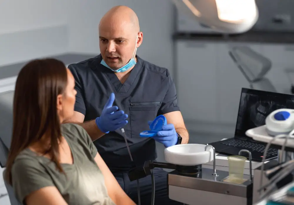
Dentists use several advanced tests to evaluate a compromised tooth’s blood supply:
- Pulse oximetry – This measures oxygen saturation in pulp blood. Lower levels indicate reduced flow.
- Laser Doppler flowmetry – A laser beam calculates microcirculation in pulp tissue based on reflected light patterns.
- Digital thermal testing – Sensors detect changes in temperature on a tooth’s surface after cold stimuli, correlating with interior blood flow.
- Electric pulp test – Applying electric current tests the vitality of nerve fibers which run alongside blood vessels. Lack of response may indicate lifeless pulp.
- Radiographs – Detailed dental x-rays show shape of root canals and bone for signs of infection and decay which impair blood supply.
- Clinical examination – Signs like pain, swelling, discoloration, fractures and mobility provide clues about blood flow status.
Once the location and extent of blood flow loss is confirmed, treatments can be tailored to try and restore circulation where possible.
Treatment options for improving tooth blood flow
If caught early, some therapies may help partially revive or preserve blood flow before it is irreversibly lost. Main approaches include:
Clearing infections, obstructions and inflammation
- Root canal therapy – Removing infected pulp and sealing the interior can resolve inflammation pressing on blood vessels.
- Root planing and debridement – Deep cleaning under the gums cleans up bacteria and tartar to improve PDL blood flow.
- Antibiotics – Prescription antibiotics like penicillin after oral surgery can prevent infections that restrict blood supply.
- NSAIDs – Anti-inflammatory pills help reduce swelling that compresses blood vessels.
Promoting circulation
- Fluoride applications – Fluoride may stimulate dentin production and strengthen enamel, supporting blood vessels.
- Occlusal guards – These protect teeth from excessive grinding forces that can strain vessels.
- Low-level laser therapy – Applying targeted laser light aims to stimulate cellular repair and increase microcirculation.
Regenerating lost pulp and blood vessels
- Pulp capping – Placing medicated dressing over exposed pulp may allow it to heal and maintain circulation.
- Stem cell therapy – Experimental use of stem cells seeks to regrow damaged pulp tissue and blood vessels.
- Scaffolds – tiny 3D-printed matrix implants could theoretically guide regeneration of vascularized pulp.
- Angiogenic growth factors – Compounds that stimulate new blood vessel formation may enable pulp revascularization.
However, current technologies still face major obstacles to fully restoring circulation inside a tooth with extensive pulp death and infection. Next we’ll examine why re-vascularization remains so challenging.
Difficulties with reestablishing tooth blood flow
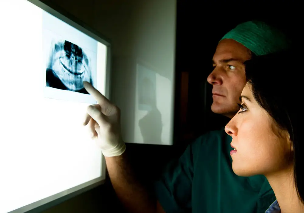
A tooth with substantial blood flow loss has often lost significant pulp tissue and sustained extensive infection. This makes it very difficult to recirculate blood due to:
- The pulp space becomes too necrotic and blocked with debris for new vessels to penetrate.
- Loss of entire networks and branches of capillaries and arterioles with few remaining connection points.
- The presence of pathogenic bacteria makes it harder for fragile new vessels to survive.
- Age-related calcification of pulp tissue further shrinks space for blood flow.
- Medical conditions like diabetes or atherosclerosis inhibit microcirculation.
- PDL and gum disease allow bacteria into dentin tubules that communicate with pulp.
- Years of inflammation makes pulp incapable of regenerating blood vessels.
- Growing new vessels risks connections with infected root canals.
- Tiny diameter of vessels sever easily and cannot be surgically reattached.
Unless infection is very minimal, it is unlikely for extensive circulation to be revived in a dead tooth. The next section examines options for managing non-vital teeth.
Treatment plans for non-vital teeth
When a tooth is deemed non-vital with irreparable blood flow, dentists must determine if it can be restored or should be removed. They assess factors like:
- Amount of remaining healthy tooth structure
- Degree of bone loss around tooth
- Presence of cracks extending below gumline
- Occlusion with opposing teeth
- Esthetic appearance of the visible crown
- Presence of active infection or abscess
- Condition of neighboring and opposing teeth
- Status of overall dental health
Based on these parameters, the main options include:
- Root canal treatment – Cleans out pulp space and seals it to prevent reinfection. Adds crown for protection.
- Root canal retreatment – Repeat root canal to resolve failed initial treatment with recurring infection.
- Extract tooth – Pulls tooth and repairs socket site. Later allows implant or bridge.
- Decoronation – Removes crown but leaves roots to preserve bone volume. No longer functional tooth.
The chosen approach depends on individual tooth prognosis and the patient’s needs. The goal is to prevent further infection and bone loss at the site.
Future outlook for tooth re-vascularization
While current therapies have limited capacity to restore circulation inside damaged teeth, emerging biomedical technologies offer hope for the future. Some promising research areas include:
- 3D bioprinting of vascularized pulp using bio-inks containing living cells.
- Further stem cell research to reliably produce pulp-like tissues integrated with blood vessels.
- Scaffolds coated with growth factors to stimulate regeneration of the pulp organ.
- Angiogenic gene therapy to increase natural angiogenesis signals.
- Nanotechnology like targeted nanoparticle delivery of regenerative compounds.
- Microsurgery and specialized stents to directly reattach severed blood vessels.
However, huge challenges remain before complex revascularization procedures become clinically feasible and effective. But the accelerating pace of medical progress predicts that creative new solutions for tooth blood flow repair may emerge within the coming decades.
Conclusion
While our current ability to restore circulation after pulp death is limited, preserving and protecting remaining blood flow is critical to saving damaged teeth. New advances in bioengineering hold future promise for revascularizing compromised teeth. But for now, prevention remains vital – regular dental visits for early diagnosis and prompt treatment of any tooth decay or trauma can help maintain robust blood supply and keep natural teeth functioning for life. With proper care, most healthy teeth should never require complex attempts at revascularization.
Frequently Asked Questions
Q: Can a dead tooth ever have blood flow return?
A: If the pulp tissue has completely necrosed, it’s likely impossible for normal blood circulation to return within the pulp chamber. But some basic flow may regenerate around the root surface.
Q: Do root canals increase blood supply?
A: Root canals aim to clear infection and inflammation from the pulp which may help partially restore flow around the root tips. But the main blood supply within the pulp itself is removed.
Q: What are signs my tooth is losing blood flow?
A: Symptoms like pain, swelling, hot/cold sensitivity, dark discoloration, negative pulp vitality tests and looseness may indicate impaired blood supply. Prompt dental exam is recommended.
Q: Can dentists put new pulp in a dead tooth?
A: Dental pulp regeneration is being researched using stem cells to grow new living pulp tissue which could theoretically be implanted into cleaned canals. But more development is needed.
Q: Is a fracture in a root canal treated tooth worse?
A: Yes, a fracture reaching below bone level in an endodontically-treated tooth has a worse prognosis due to lack of blood supply. It becomes at higher risk for infection and complications.

