What is tooth decay and how does it occur?
Tooth decay, also known as dental caries or cavities, occurs when acids dissolve the mineral content of teeth, causing holes or lesions to form. It is caused by oral bacteria like Streptococcus mutans and Lactobacillus that colonize on tooth surfaces in a sticky, colorless biofilm called plaque.
These bacteria metabolize and ferment simple carbohydrates from the food we eat, particularly refined sugars and starches, into acids. The most common acid produced is lactic acid, which can create an acidic environment in the mouth with a pH below 5.5. At this pH level, the enamel, which is the outermost layer of the tooth, begins to demineralize and dissolve, causing cavities.
Demineralization from acid attacks causes minerals like calcium, phosphate and carbonate to leach out of the enamel rods. This causes microscopic pores to form and the enamel to weaken. The acids also irritate the gums, making them tender, swollen and more prone to periodontal disease.
If this demineralization continues over time, these pores can accumulate and spread to form a cavity. The cavity can then continue into underlying dentin and cementum layers of the tooth. Dentin contains thousands of microscopic tubules leading to the nerve-rich pulp inside the tooth. As the decay reaches this layer, it can cause sensitivity and pain. The inner pulp tissue becomes inflamed from bacteria and eventually dies if decay progresses untreated.
How does a root canal help stop tooth decay?
Root canal treatment is recommended when tooth decay has damaged or infected the soft pulp tissue inside the tooth. It involves removing the diseased pulp, disinfecting the hollowed-out root canals and sealing them to prevent further infection.
Here are the key steps:
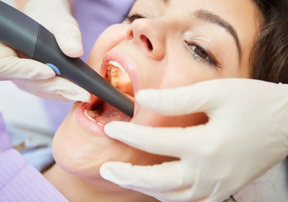
- The dentist administers local anesthesia and makes an opening through the enamel and dentin layers to access the pulp chamber.
- The inflamed or necrotic pulp tissue is removed using small dental instruments. The root canals are then cleaned and shaped using irrigating solutions. This flushes out dead tissue remnants and bacteria from the intricate canal anatomy.
- The entire root canal system is then disinfected with antimicrobial rinses. This eliminates remaining bacteria.
- The clean, hollowed out canals are filled and sealed with an inert rubbery material called gutta percha. This prevents reinfection of the tooth.
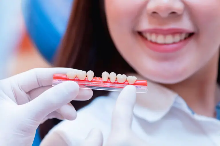
- A temporary filling is placed over the access cavity and a final restoration like a dental crown is fabricated to seal the tooth.
By removing infected pulp tissue and cleaning out bacteria, a root canal stops the decay process and prevents further demineralization of tooth structure from rampant cavities. It conserves the tooth so that restorative options like crowns can be placed.
Why does decay still occur after a root canal procedure?
Though a root canal treatment stops an active carious infection, restored teeth may still be at risk of developing new cavities for several reasons:
- Remaining bacteria: The intricate root canal system has tiny ramifications and anatomical complexities where bacteria could potentially still lurk after disinfection. If these leftover microorganisms proliferate, they can leak out through the dentinal tubules and trigger new caries formation on the external tooth surface.
- Microscopic tubules: Dentin has thousands of microscopic channels leading from the pulp chamber to the enamel called dentinal tubules. They contain fluid and organic matter. After a root canal treatment, these tubules remain patent and can transfer bacteria from the contaminated root canal to the outer enamel surface where acids are produced.
- Cracks and defects: Root canal treated teeth become more brittle and prone to cracks due to loss of their nerve and blood supply. Cracks create sheltered environments for bacteria and acids to congregate, increasing decay risk.
- Residual caries: If all infected and softened enamel and dentin are not completely removed before placing the final restoration, any remaining decayed tissue can continue to advance and undermine the restoration.
- Open restorations: Leaking fillings and dental restorations allow oral bacteria, fluids and food debris to seep in. Bacteria can then colonize on the tooth subsurface and cause recurrent decay.
- Reduced salivary flow: Some root canal medications reduce saliva flow temporarily. This decreases the neutralizing, antibacterial and remineralizing benefits of saliva, increasing vulnerability to new cavities.
How likely is recurrent decay after a root canal procedure?

According to dental research, the likelihood of new decay developing on a tooth after root canal treatment ranges widely:
- One study found 10-year success rates of root canal treated teeth with posts and crowns to be between 89-93%. This suggests a 6-10% incidence of complications like recurrent decay within a decade.
- Another study reported that nearly 50% of root filled molar teeth developed new caries within 4 years of treatment. The rate was higher for molars needing retreatment compared to first-time treatments.
- Several reviews estimate that depending on factors like oral hygiene, eating habits, saliva flow and type of final restoration, the chances of recurrent decay range from 2% up to 48% over varying periods.
- Data indicates the risk is highest within the first year after root canal treatment, especially for molars and premolars. After 5 years, decay rates tend to plateau if the seal of the final restoration remains intact.
So while root canals do not completely eliminate chances of new decay, the risk can be minimized by good oral hygiene, regular dental visits for early caries detection,strict control of diets with refined carbohydrates and sugars, and properly sealed final restorations.
Can tooth decay occur under dental crowns placed on root canal treated teeth?
Dental crowns fully encapsulate the visible portion of a tooth down to the gum line. However, decay can still occur in the uncovered root area below the margins of the crown.
Some ways this can happen include:
- Defective crown margins: Poorly contoured or open crown edges allow bacteria, fluids and food debris to seep underneath the crown. This lets decay initiate on the unprotected root surfaces.
- Improper crown fitting: Ill-fitting crowns have microscopic gaps between the edge and tooth. Oral bacteria accumulate in these spaces and cause decay.
- Compromised dental cement: Weakened cement allows fluid movement and bacterial entry under the crown that can start root decay.
- Exposed dentinal tubules: Where the root area is devoid of protective cementum covering, open dentinal tubules allow rapid bacterial invasion from the crown edge to the root surface to spur decay.
- Loss of periodontal attachment: Severe gum recession from periodontal disease exposes more of the root area to oral bacteria. This increases susceptibility to root decay under the crown margins.
- Previous root defects: Existing root caries, cracks or defects not completely treated before crown placement can worsen under the crown due to bacterial ingress.
- Poor crown contours: Overhanging or poorly shaped crown edges prevent proper plaque removal, promoting decay in those stagnant areas.
Therefore, while crowns protect the visible coronal portion of root canal treated teeth, their margins and surrounding root surfaces remain vulnerable. Maintaining good oral hygiene and regular dental visits can help detect and treat such recurrent decay in the early stages before extensive damage occurs under the crown.
What are some warning signs of tooth decay under dental crowns?

Some warning signs that may indicate an underlying cavity or decay developing under a dental crown include:
- Tooth sensitivity, especially to hot or cold foods and drinks. This suggests inflammation of the tooth pulp by progressing decay.
- Pain or discomfort when biting down or when releasing the bite. This is due to pressure on decayed areas irritating the tooth’s nerve.
- A foul, sulfur-like odor emerging from the gums around a crowned tooth, signaling infection.
- Persistent bad breath or altered taste sensations that don’t resolve with brushing. This may arise from bacterial accumulation and gases escaping from decaying areas under the crown.
- Gum inflammation – swollen, red or bleeding gums near the crowned tooth. Advancing decay elicits an inflammatory response.
- Changes in the color of the gums around the crown, like redness or blackening. Discoloration can indicate spreading decay.
- Difficulty removing food debris from below the crown. Trapped particles accelerate decay at the crown-tooth junction.
- A visible gap forming between the crown edge and tooth surface, signalling deteriorating cement.
- A loose crown that moves slightly on biting indicates loss of crown retention from decaying tooth structure.
- A change in the fit of the crown in the bite over time as decay alters dimensions.
Seeing a dentist promptly for evaluation of any such symptoms allows early intervention before the decay worsens extensively under the crown.
How is tooth decay diagnosed and treated if found under a dental crown?
If tooth decay is suspected under an existing crown, the dentist will first clinically examine the tooth for any signs like discoloration or gaps at the crown edge. Sensitvity and percussion tests may be done to check for inflammation in the tooth pulp. The supporting bone will also be evaluated for any deterioration.
If clinical tests are positive for potential decay, radiographic imaging like bitewing x-rays or cone beam scans will be taken to visualize the extent of damage. The images may show definitive lesions or dark areas indicating advanced decay.
If decay is confirmed under the crown, several treatment approaches are possible depending on its severity:
- If the decay is minimal and shallow, just the crown can be removed, decay excavated, and the crown recemented after placing an updated base or liner.
- If the decay is moderate but has not reached the pulp, the crown can be sectioned and tooth structure rebuilt with an adhesive filling before fabricating a new crown.
- For extensive damage involving the pulp, the tooth may require root canal retreatment followed by a post and core buildup and new crown.
- Extraction may be necessary if the tooth decay is too severe and the remaining tooth structure cannot support a restoration. Replacement options like dental implants can then be considered.
- If the patient is asymptomatic, a no treatment approach with close monitoring every 3-6 months may be appropriate for minimal decay.
With timely diagnosis and intervention, many teeth with recurrent decay under crowns can be successfully retreated and restored to full function. Maintaining diligent oral hygiene and regular dental visits are key to minimizing such post-treatment complications.
Frequently Asked Questions
1. Why do some root canal treated teeth still decay?
Some reasons root canal treated teeth may still decay include:
- Residual bacteria left in microscopic areas of the tooth
- Exposure of dentinal tubules allowing bacterial transfer
- Cracks and defects in the treated tooth
- Poor oral hygiene allowing plaque buildup
- Inadequate caries removal prior to final restoration
- Reduced salivary flow decreasing natural defenses
2. How long do crowns last on a root canal treated tooth?
With good oral care, crowns on root canal treated teeth can last 10-15 years or longer. However, defective crown margins, gum recession, strong biting forces and recurrent decay can shorten survival times.
3. Can X-rays detect a cavity starting under a crown?
Yes, dental x-rays like bitewings and periapicals can detect decay starting as a small radiolucent spot under a crown if the decay has progressed enough to demineralize the enamel and dentin. However, early decay may not always show up.
4. Is a root canal absolutely necessary if decay is found under a crown?
Not always. For minor decay just under the crown margin, a root canal may be avoided by removing the crown and filling the defect. However, extensive decay reaching the pulp necessitates root canal treatment. The dentist will determine this based on the decay depth and severity.
5. What is the success rate of root canal treatment with a crown?
The reported success rate of initial root canal therapy followed by placement of a full coverage restoration like a crown is very high, in the range of 86% to 97% based on various studies. The success depends on many factors like bone support, oral hygiene, saliva flow and choice of restorative treatment.