Teeth are secured in the jawbone for strength and function
Teeth are made to last a lifetime. Their structure and position in the jaw gives them the strength to withstand the forces of biting and chewing. This is why they can be so difficult to remove.
Teeth have two parts – the crown that is visible above the gums, and the root below the gums embedded in the jawbone. The root provides stability and helps anchor the tooth in place. It is held tightly within the bony socket by a fibrous attachment called the periodontal ligament. This ligament acts like a sling to keep the tooth from moving around.
Tooth anatomy for strength
The tooth itself is made of 4 tissues that give it hardness and durability:
- Enamel – The outermost layer that covers the crown. It is the hardest substance in the body.
- Dentin – Bony tissue layer under enamel that makes up most of the tooth.
- Pulp – Soft tissue at the center containing blood vessels and nerves.
- Cementum – Bonelike tissue covering the root that anchors it to the jaw.
This combination of hard mineralized tissues and fibrous attachments makes natural teeth strong and difficult to dislodge or extract.
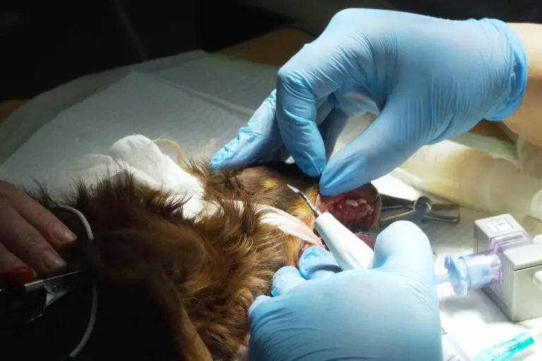
Extraction process places force on the tooth

During an extraction, the dentist must loosen the tooth from the socket and break the periodontal ligament attachments. This requires controlled force applied in the right direction.
There are two main extraction techniques:
- Simple extraction – Used when the tooth is visible and can be grasped with forceps. Outward force loosens the socket attachments.
- Surgical extraction – Used when the tooth is broken, decayed or impacted. Some bone may be removed and the tooth is separated into pieces for removal.
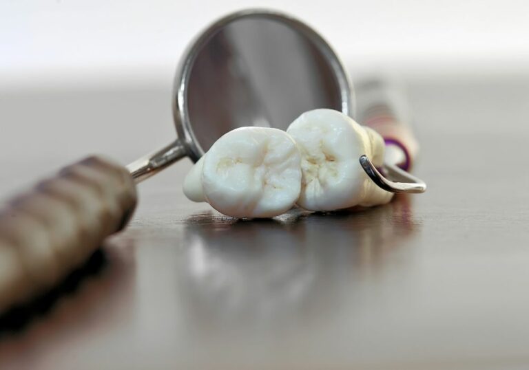
The amount of force depends on root shape and how well anchored it is in the bone. Molars can be especially difficult due to multiple curved roots.
Extraction process step-by-step
Here is the extraction process that shows where force must be applied:
- Numbing – Local anesthetic blocks pain and widens space around the tooth.
- Loosening – Tools are used to loosen ligament attachments and widen the socket.
- Rocking – The tooth is rocked back and forth to break ligament fibers.
- Removal – Finally, the tooth is ready to be removed from the socket.
This sequence places controlled stress on the extraction site. The tooth roots resist being dislodged, requiring significant pressures.
Factors that increase tooth removal difficulty
Many issues can make a tooth extraction more challenging, such as:
- Tooth position – Impacted, twisted or awkward angles require intricate maneuvers.
- Root anatomy – Long, curved or excessively divergent roots resist removal.
- Dense bone – Very hard, thick jawbone restricts loosening the tooth.
- Previous dental work – Crowns, bridges or implants complicate the procedure.
- Patient cooperation – Anxiety makes patients move or clench their jaws.
These factors mean the dentist must work incrementally with specialized instruments to extract the tooth while protecting the surrounding bone.
Tooth extraction difficulty scale
Teeth can be ranked by anticipated extraction difficulty:
| Tooth Type | Extraction Difficulty |
|---|---|
| Single rooted | Simple |
| Multi-rooted | Moderate |
| Impacted/Fused | Very Difficult |
As more resistance is met, the dentist must apply greater forces from just the right angle to complete the extraction successfully.
Options for removing difficult teeth

For teeth that do not respond to conventional extraction techniques, oral surgeons have other tools to facilitate the process:
- Elevators – Flat metal tools for loosening periodontal ligament fibers.
- Luxators – Curved instruments to loosen teeth and widen the socket.
- Physics forceps – Uses levers and principles of physics to improve grip and pull.
- Surgical burs – Rotary drills to remove obstructing bone around the tooth.
- Sectioning – Cutting a multi-rooted tooth into pieces for easier removal.
These advanced options allow the surgeon to wedge, splinter and cut the tooth until it can be detached. The goal is to remove it as atraumatically as possible.
When extractions are too risky
In some cases, an extraction is deemed too difficult or prone to complications. The tooth may be left in place and monitored if:
- Severe infection is present
- It may fracture the jawbone during removal
- Patient health will not permit surgery
This avoids trauma, but the tooth should continue to be evaluated.
Extraction complications are still possible
Even with advanced techniques, extractions carry risks, including:
- Bone fragments – Small fragments of bone can break off in the socket.
- Sinus tear – Upper teeth are close to the sinus and could puncture this area.
- Nerve injury – The inferior alveolar nerve runs near the lower teeth and may be bruised or damaged.
- Jaw fracture – Applying excessive torque could fracture the jawbone.
- Dry socket – Slow healing after extraction can leave exposed bone painful and at risk of infection.
Proper recovery care is needed after an extraction to prevent these issues which could require further treatment.
Signs of extraction complications
Patients should watch for the following problems and contact their dentist if they occur:
- Severe pain that steadily worsens
- Significant swelling inside or outside the mouth that expands
- New numbness or altered sensations in the face, teeth or mouth
- Difficulty opening the mouth or jaw stiffness
- Exposed bone within the socket with intense throbbing pain
Seeking prompt care can minimize the impact of any complications.
Maintaining long term tooth and bone health

Once a tooth is extracted, steps should be taken to preserve the surrounding bone and prevent neighboring teeth from shifting. This may include:
- Socket preservation – Materials are placed in the socket to encourage bone regrowth. This prevents bone loss.
- Bone grafts – Existing bone can be augmented to strengthen the site for implants or bridges.
- Ridge expansion – Inadequate bone width can be expanded for replacement teeth.
- Tooth replacement – Options like bridges, partials or implants prevent tipping of adjacent teeth into the space.
With proper planning and restorative treatment, extraction defects can be corrected and a fully functioning mouth maintained.
Tooth replacement timeline after extractions
Here is a general timeline for tooth replacement procedures following one or more extractions:
| Procedure | Timing after extractions |
|---|---|
| Socket preservation | Immediately |
| Bone graft | 2-3 months healing |
| Dental implant | 3-6 months healing |
| Bridge placement | 1-2 weeks healing |
| Partial denture | 2-4 weeks healing |
Consultation with your dentist will determine the appropriate treatment schedule for your situation.
Conclusion
Teeth are tenaciously secured in the jaw by their mineralized structure and the holding power of the periodontal ligament. These attachments make them difficult and technique-sensitive to remove. Dentists utilize complex maneuvers and tools to loosen teeth and draw them successfully from their sockets. Even with proper precautions, extractions still carry risks of complications which must be managed promptly. Careful planning can restore the area for long term dental health after a tooth is extracted.
Frequently Asked Questions
Why are some teeth easier to extract than others?
The shape and number of roots determines how readily a tooth can be extracted. Single rooted teeth like incisors and canines have a conical single root that tapers to an apex. These slide out of the socket fairly easily with simple rocking motions. In contrast, molars have multiple divergent roots that branch and curve in different directions. The complex anatomy requires cutting through gum tissue to remove bone and section the tooth for extraction in pieces.
Is a “simple” extraction still difficult?
Even a simple extraction is still challenging due to the tooth’s bony composition and the forces needed to disrupt its socket attachments. However, simple extractions generally involve front teeth with straight single roots that can be elevated in one piece. This takes less time and surgical intervention than removing teeth with multiple curved roots or unusual positioning in the jaw.
What teeth are the most difficult to extract?
Third molars or wisdom teeth are usually ranked as the most difficult extractions. Their position at the very back of the mouth next to vital nerves and sinuses along with their multi-rooted anatomy and tendency for impaction makes them challenging to remove. Canines can also offer resistance due to their single large root and location in the “bite zone.”
Can smoking affect the difficulty of extractions?
Yes, smoking negatively impacts tooth extractions and healing. The chemicals in tobacco can cause bone loss and make the bone that remains harder in consistency but weaker in structure. Smokers tend to have increased rates of healing complications. Dentists often recommend smokers cease tobacco use for several weeks before and after planned extractions.
Do dentists ever have to stop in the middle of an extraction?
It is possible that an attempted extraction may fail or need to be halted mid-procedure. This can occur if the tooth fractures within the jawbone, endangers a nerve or artery, or cannot be completely separated from its socket attachments. Temporary stopping measures may be taken while further imaging and planning is done to revise the approach before attempting the extraction again.