Teeth and bones share common developmental origins, arising from oral ectoderm and cranial neural crest cells. However, key differences in their adult composition and structure lead to very different potential for self-healing and regeneration. While bone tissue can completely regenerate and heal itself, teeth have very limited natural healing abilities.
Key Structural Differences Between Teeth and Bone
Teeth and bones have distinct architectural designs that influence healing capacity:
Tooth Anatomy
- Enamel – Hardest substance in body, mineralized epithelial cells. Acellular.
- Dentin – Softer than enamel but still rigid, contains some odontoblast cells.
- Pulp – Soft tissue core containing nerves, blood vessels, lymphatics, and cellular elements.
- Cementum – Bonelike calcified substance covering roots, contains cementocytes.
- Periodontal ligament – Suspends and supports teeth in jawbone, some cellular elements.
Bone Anatomy
- Compact bone – Hard external layer, dense concentric lamellae around central canals.
- Spongy bone – Web-like trabecular networks with marrow-filled voids.
- Periosteum – Outer membrane covering bone, contains nerves, blood vessels, lymphatics.
- Endosteum – Lines medullary cavity, assists bone remodeling.
- Blood vessels – Penetrate through Volkmann’s canals to nourish bone cells.
This table compares tooth and bone anatomy:
| Tissue | Location | Function |
|---|---|---|
| Enamel | Outer layer of crown | Protective hard shell |
| Dentin | Under enamel | Supportive & protective |
| Pulp | Center | Nerves & blood supply |
| Cementum | Covers roots | Anchors to bone |
| Periodontal Ligament | Around roots | Suspension in socket |
| Compact Bone | Outer shell | Structural support |
| Spongy Bone | Inner trabeculae | Metabolic activity |
| Periosteum | Outer surface | Covers and nourishes |
| Endosteum | Lines medullary cavity | Remodeling agent |
| Blood Vessels | Canals | Nutrient supply |
The varied tissues comprising teeth and bone provide different architectural support suited to their distinct functions.
Cellular Composition Differences
The living cellular composition of bones and teeth diverge as well:
Bone Cells
- Osteoblasts – Bone forming cells
- Osteoclasts – Bone resorbing cells
- Osteocytes – Mature bone cells
These perform remodeling through osteons, tuning bone shape/strength.
Tooth Cells
- Odontoblasts – Form dentin, stay viable in pulp
- Ameloblasts – Form enamel, lost after development
- Cementoblasts – Form cementum, remain as cementocytes
No cells in bulk of enamel or dentin for remodeling.
Stem Cell Differences
- Bone marrow contains mesenchymal stem cells that can form osteoblasts.
- Dental pulp contains undifferentiated mesenchymal cells with limited differentiation capabilities.
This table outlines key cellular distinctions:
| Bone Cells | Role | Tooth Cells | Role |
|---|---|---|---|
| Osteoblasts | Bone forming | Odontoblasts | Dentin forming |
| Osteoclasts | Bone resorbing | Ameloblasts | Enamel forming |
| Osteocytes | Bone maintenance | Cementoblasts | Cementum forming |
| MSCs | Generate osteoblasts | Dental stem cells | Minimal roles |
In summary, bones possess active living cells to dynamically remodel tissue, while teeth lose cells needed for remodeling.
Structural Component Differences
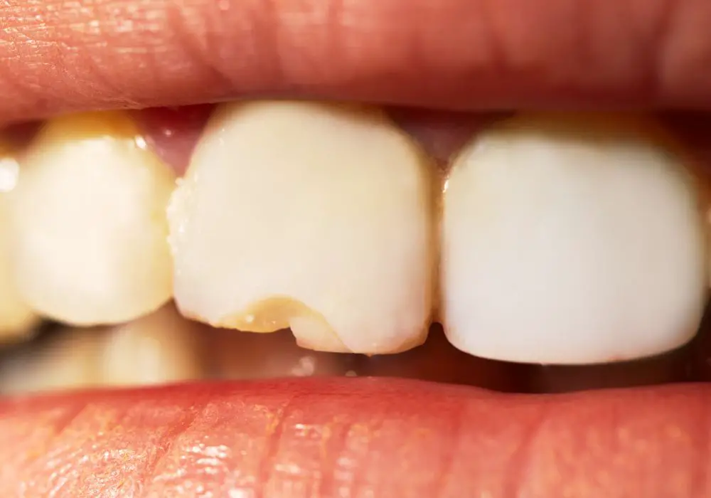
Teeth and bones differ in their fundamental biomaterials, impacting healing:
Bone Matrix
- Collagen fibers – Flexible protein comprising 30% of bone matrix.
- Hydroxyapatite – Mineral crystals comprising 70% of bone matrix.
Collagen turnover by osteoblasts facilitates bone flexibility and self-repair.
Tooth Matrix
- Dentin – 70% hydroxyapatite, 20% collagen matrix.
- Enamel – 96-98% hydroxyapatite, no collagen.
Tooth mineralization prevents collagen turnover for self-repair.
This table compares structural composition:
| Material | Bone Matrix | Tooth Matrix |
|---|---|---|
| Collagen | 30% by weight | ~20% in dentin only |
| Hydroxyapatite | 70% by weight | 96-98% in enamel |
The lack of collagen and increased mineralization in tooth structural components limits remodeling.
Innervation Differences
Nerves interact differently with bone versus teeth:
- Sensory nerves in bone are sparse but cover periosteum surfaces.
- By contrast, tooth pulp is densely innervated to support sensation.
- Autonomic nerves control bone blood flow but are minimal in teeth.
- Neuropeptides aid bone remodeling but are lacking in teeth.
Thus, bones contain more integrative nerves to coordinate healing responses than teeth.
Vascular Supply Differences
Bone tissue relies on extensive blood supply:
- Arteries, veins, and capillaries penetrate compact bone through Volkmann canals.
- Spongy bone is infiltrated by red marrow containing blood vessels.
By contrast, only the pulp core of the tooth receives any blood supply, through the apical foramen. The bulk of mineralized tooth tissue is avascular.
Robust vascular supply is critical for delivering healing cells/factors to bone. Teeth lack this benefit.
Developmental Differences
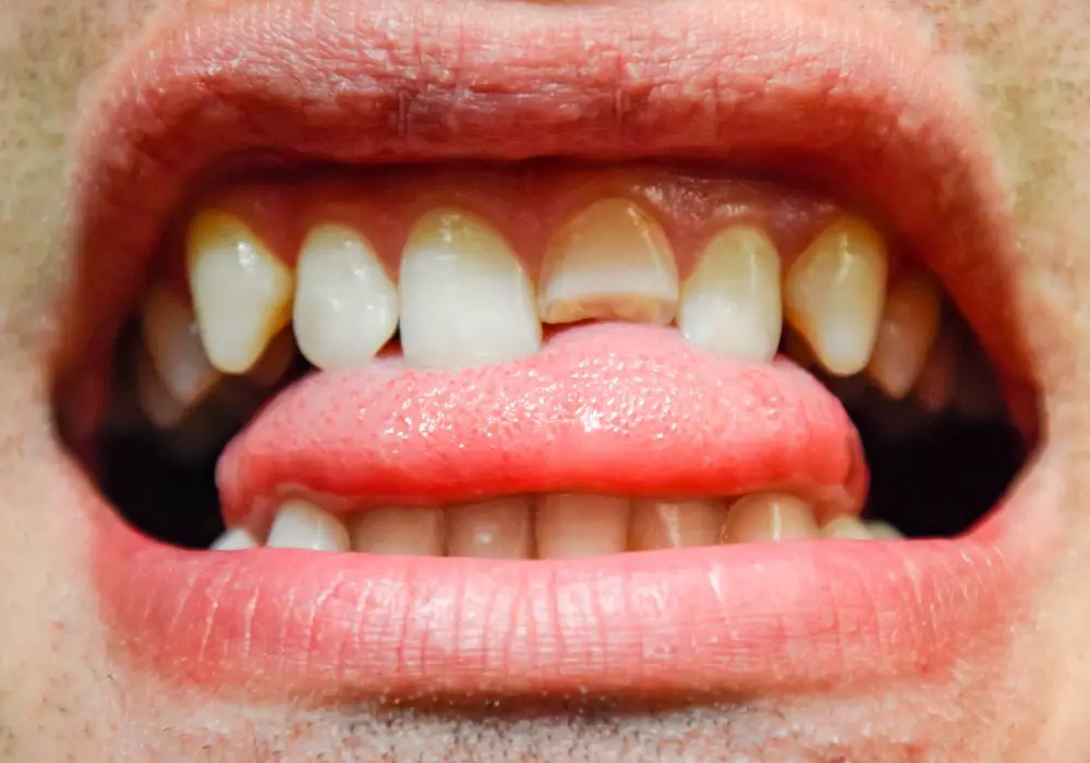
Despite some common embryological origins, bone and tooth development diverges:
Bone Development
- Most craniofacial bones formed via intramembranous ossification.
- Long bones formed by endochondral ossification.
- Involves coordinated differentiation of ossification centers.
Tooth Development
- Controlled by sequential and reciprocal epithelial-mesenchymal interactions.
- Establishes spatial patterning for complex multi-tissue morphogenesis.
- Guided by molecular signals like BMPs, FGFs, SHH.
These distinct developmental mechanisms create optimized functional structures.
Summary of Key Differences
In summary, bones and teeth diverge in:
- Architectural structure
- Living cellular content
- Biomaterial composition
- Innervation patterns
- Vascular supply
- Developmental processes
These factors critically determine natural healing capabilities.
Impacts on Healing Potential
The impacts of these differences on healing are clear:
Bone Healing
- Bone is living tissue with regenerative cells (osteoblasts, osteoclasts, MSCs).
- Robust blood supply delivers nutrients and healing factors.
- Collagenous matrix can be remodeled and reformed.
- Developmental processes can be recapitulated to regenerate bone.
Tooth Healing
- Non-living mineralized tissues lack regenerative cells except in the pulp.
- Minimal blood supply prevents delivery of healing factors.
- Rigid hydroxyapatite matrix cannot be remodeled.
- Developmental processes cannot be fully replicated to regrow teeth.
Consequently, bones have intrinsically greater healing potential than teeth. Although treatments can aim to improve tooth regeneration, intrinsic constraints remain.
Clinical Treatment Implications
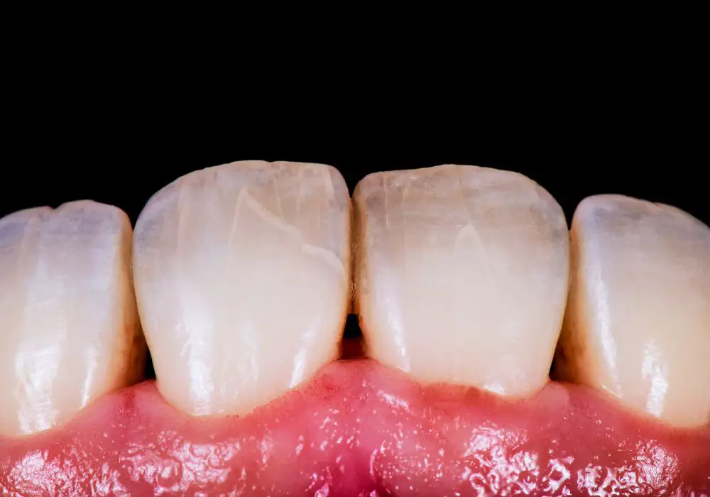
These fundamental differences have profound impacts on clinical treatment approaches:
Bone Fracture Treatment
- Casting stabilizes bone segment alignment for optimal natural healing.
- Surgical fixation with plates or screws can also promote healing.
- Bone grafts or growth factors are sometimes used to further enhance regeneration.
Dental Cavity Treatment
- Decayed tooth structure cannot regenerate on its own.
- Restorations like fillings or crowns are required to replace lost tissue.
- Major damage may require artificial implants or bridges since regeneration is not possible.
Understanding the inherent constraints on tooth healing guides selective use of reparative therapy rather than relying on the body’s regenerative capacity.
Future Directions
Advancing research aims to enhance tooth healing potential:
- Stimulating natural tooth regeneration using biomaterial scaffolds and stem cell therapy.
- Developing methods to induce tooth tissue de- and re-mineralization.
- Modulating developmental signaling pathways to regrow teeth.
- Bioengineering whole tooth units for transplantation.
While total biological tooth renewal remains challenging, incremental progress continues toward this goal.
Conclusions
In summary:
- Profound differences exist between bones and teeth in structure, cells, matrix, nerves, and blood supply.
- These lead to bones having intrinsic regenerative capacity while teeth have very limited ability to self-heal.
- Clinical treatments reflect this dichotomy, focusing on facilitating bone regeneration while replacing damaged tooth tissues.
- Advances in regenerative medicine and developmental biology may one day allow teeth to recapitulate bone’s healing powers.
Recognizing the biological basis for constraints on natural tooth healing is key to developing improved therapeutics aimed at enhancing regenerative capacity. This knowledge guides current practice and future directions in restorative and regenerative dentistry.
Frequently Asked Questions
Why can’t teeth regenerate like bones can?
The key factors limiting tooth regeneration compared to bone include: 1) Lack of living cells to drive remodeling in mineralized tooth tissues; 2) Absence of collagenous matrix that can be reformed; 3) Insufficient vascular supply to bring regenerative cells/factors; 4) Inability to recapitulate the developmental processes that originally formed the tooth.
What cells could help regrow enamel or dentin?
Enamel cannot regenerate since ameloblasts are lost after tooth development. Odontoblasts remain viable in the pulp to maintain dentin, but cannot substantially reform damaged dentin. Stem cell therapy may eventually help regrow dentin by differentiating new odontoblast-like cells.
How does the blood supply differ between bone and teeth?
Bone has an extensive blood supply with arteries/veins penetrating through Volkmann’s canals to reach all areas. Teeth only have blood vessels in the pulp, leaving the bulk of mineralized tissue with no direct blood supply. This limits delivery of nutrients/healing factors in teeth.
Why can fractures heal but cavities cannot?
Bone fractures involve damage to living tissue that contains regenerative cells like osteoblasts that can rebuild the bone. Cavities affect acellular enamel/dentin that lack ability to regenerate. Only artificial restorations can replace the cavity, rather than relying on natural healing processes.
What advances are needed for biological tooth regeneration?
Key advances needed include: identifying optimal stem cell sources, development of scaffold biomaterials, recapitulating developmental signaling pathways like Wnt, BMP, and FGF, establishing vascular supply, and integrating biomimetic approaches into regenerative strategies.
