Human teeth are composed of remarkably sturdy tissues designed to persist for decades of constant use during life. But teeth also have the capacity to endure for remarkably long periods after death as well, essentially becoming fossilized inside the jawbones and skulls of skeletal remains. While all other soft tissues decay away, teeth can linger in place, providing a lasting record of long-deceased individuals. But what exactly accounts for the ability of teeth to withstand degradation and remain embedded within skeletons for so long? There are a number of biological factors that enable teeth to endure while bones and soft tissues disappear.
Tooth Structure and Composition
Teeth are comprised of multiple mineralized hard tissues specialized for mechanical stability and wear resistance. The outermost layer of a tooth is the enamel, which covers and protects the underlying softer dentin and inner pulp.
Enamel
Enamel is made up of tightly packed hydroxyapatite crystals that form an incredibly hard, dense, durable tissue. Hydroxyapatite is a highly calcified crystalline form of calcium phosphate, akin to geological apatite found in fossils. The unique hexagonal prism-like structure of enamel crystals provides enhanced physical strength. Enamel contains the highest mineral content of any tissue in the body at 96-98% mineral salts with only 2-4% organic matter and water. This extraordinarily high mineral concentration makes enamel more resistant to decomposition than any other tissue.
Enamel thickness varies from 2.5 mm on molars to mere micrometers at the cervical margin where the crown meets the root. The crystalline structure and thickness provides protection and wear resistance during a lifetime of mastication. But those same properties also allow enamel to persist relatively unchanged for extraordinary lengths of time after death. While enamel can chip, crack, or wear over thousands of years, it resists chemical and bacterial degradation.
Dentin
Dentin makes up the bulk of a tooth, situated beneath the enamel and surrounding the inner pulp chamber. It contains 70% mineral salts in the form of hydroxyapatite crystals embedded in a 20% organic collagen matrix with 10% water content. This lower mineral but higher collagen content makes dentin more flexible and fracture resistant than enamel. However, it is still quite hard and durable. The properties of dentin also lend itself to long-term preservation in skeletons.
Cementum
Cementum is a specialized bony substance covering the tooth root that anchors teeth to the alveolar bone through the periodontal ligament. It attaches collagenous Sharpey’s fibers to cementum and jawbone matrix for stability. Cementum resists compression forces on teeth during chewing and grinding. It can remain on embedded tooth roots after death for some time but tends to slough off ancient teeth as bone matrix disappears.
Pulp
Pulp occupies the hollow inner chambers of teeth and contain blood vessels, nerves, and connective tissue. The cellular contents and structure of pulp rapidly breakdown after death through autolysis and bacterial decay once the protective enamel shell is compromised. However, the pulp chamber shape often remains intact long after its soft contents are gone.
In life, the combination of highly rigid calcified enamel and dentin with softer organic pulp and attached periodontal membranes allows teeth to withstand immense compressive chewing forces and remain firmly anchored in socket bony tissue without fracturing. After death, the pulp tissue decomposes but leaves behind the incredibly durable enamel and dentin minerals accounting for 97% of total tooth volume. This substantial residual mineral content is why most of a tooth’s crown and root structure can endure for so long after all cells, proteins, and soft tissues have disappeared.
Lack of Moisture Decelerates Decay
The conditions surrounding decomposition have a major influence on tooth retention over time. Teeth are much more prone to post-mortem loss from skeletons in humid environments than in hot, arid conditions. Moisture enables bacteria and enzymes to breakdown and dissolve tissues through putrefaction.
Bacterial degradation is accelerated by ambient moisture, which allows microbes to thrive and produce collagen-digesting enzymes that can slowly dissolve dentin. Bacterial hydrolysis of dentin’s organic matrix helps loosen teeth from deteriorating sockets. Teeth may literally decay out of the jawbone under active bacterial attack when moisture is present.
Alkaline hydrolysis from anaerobic bacteria can also slowly dissolve and liquefy hydroxyapatite minerals when moisture is abundant. The dissolution is enhanced by bacterial acids and diffusion of hydroxyl ions into apatite crystals.
Conversely, hot and dry conditions characteristic of deserts or protected tombs essentially mummify remains and teeth through desiccation. When a body completely dries out, bacterial activity and chemical reactions are halted. Tooth decay depends heavily on moisture to enable microbial and enzymatic processes. Aridity retards decomposition and leaves structures intact. So dryness greatly enhances the preservation potential of archaeological teeth over timespans of centuries and millennia.
The best examples of long-enduring teeth come from Egypt and other hot desert regions where moisture is extremely limited. Teeth have persisted while essentially all soft tissues vanished from mummified remains. Skeletal hands sometimes still grasp teeth because surrounding bones decomposed quicker in humid burial soil.
Jawbone Structure and Tooth Attachment
Teeth are not simply floating in soft tissue but are firmly implanted into the rigid maxillary and mandibular bones of the upper and lower jaw. The bony sockets, known as alveoli, help structurally support and contain the roots except for some exposed crown area. The periodontal ligament reinforces the tooth roots by attaching the cementum covering to adjacent socket bone via collagenous Sharpey’s fibers. This anchoring system combined with tight crown fit resists displacement of teeth even as surrounding alveolar bone deteriorates over time.
The upper maxilla bones contain more porous and delicate trabecular bone matrix which decomposes quicker than the denser mandibular jawbone. Maxillary molar roots are shorter with less surface area attached by periodontal fibers. So the upper dentition relies more on gingival attachments and is slightly more prone to loss. The thicker mandible and extra root surface area and fiber attachments of lower molars keep them firmly lodged. Mandibular teeth often remain many years after maxillary arch integrity fails.
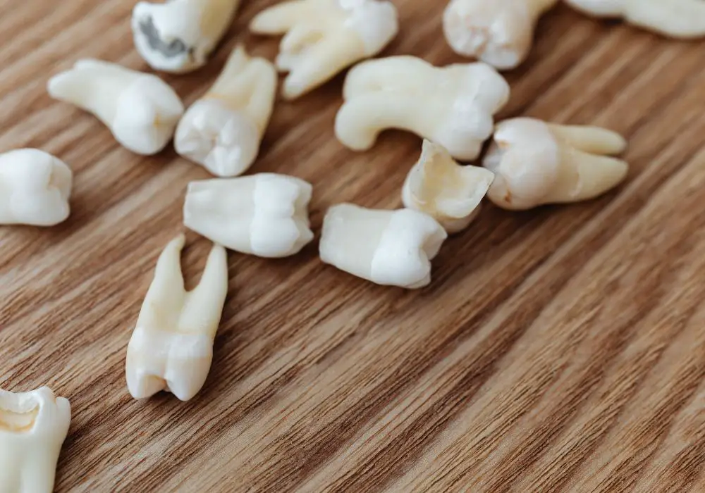
The number and orientation of roots also plays a role. Multi-rooted mandibular molars have several periodontal fiber bundles securing each root branch. The curved, diverging roots help lock these teeth in place. Small, single-rooted anterior teeth are inherently less stable.
So the anchoring bone, periodontal tissues, and root structure retains teeth in precise positions for a significant time after death even as the surrounding maxilla and mandible bones resorb. Tooth crowns prevent easy upward displacement from socket walls. This structural system persists allowing recognition of original tooth positions and occlusion long after the body is skeletonized. Eventually though, the degraded alveolar bone can no longer support complete dentition.
Tooth Type Durability and Loss Patterns
Not all teeth have the same size, shape, root structure, and composition. So certain teeth tend to remain embedded in jaw bones for longer durations after death than others which are more prone to dropping out as ligaments detach and bone resorbs.
Molars
Molars are the largest type of teeth with the most durable physical attributes conducive for long-term in situ preservation. They have:
- Largest proportion of highly resistant enamel
- Greater depth and area of enamel coating
- Thickest layer of enamel (up to 2.5 mm)
- Multiple divergent roots (2 on premolars, 3 on molars)
- Broad surface area of periodontal ligament attachment
- Tighter alveolar socket fit from rounded roots
- Vertical directed forces during chewing
This combination of extensive enamel and multiple wide-spreading roots anchored into a thick mandible body allows most molars to remain intact potentially for thousands of years. Even when cracking and wear eventually occurs, their initial stability gives molars the best chance of persisting in situ until ancient times relative to other tooth types.
Incisors and Canines
Anterior incisors and canines conversely have a much higher likelihood of postmortem loss. Contributing factors include:
- Smaller amount and thickness of enamel
- Single conical root form
- Less socket contact area
- Primarily horizontal chewing forces
- Location in fragile maxilla
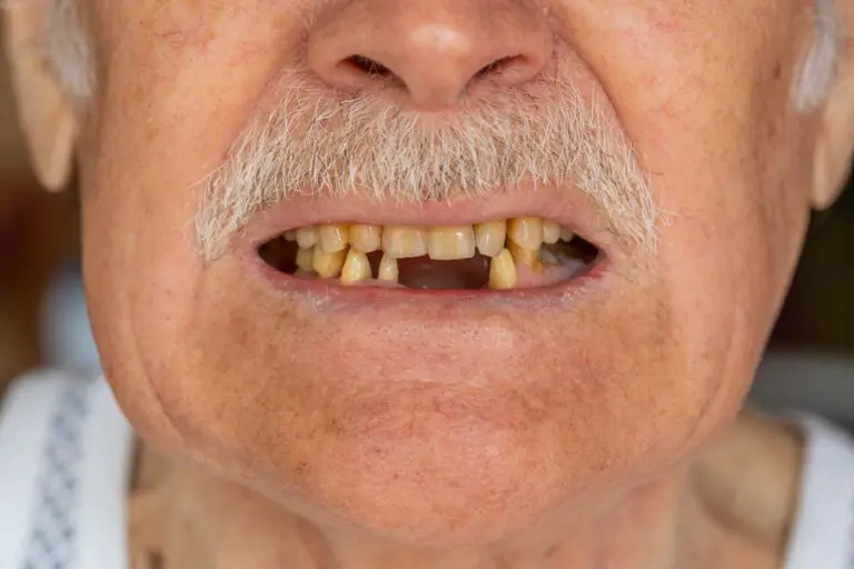
The single slender root provides little resistance to displacement compared to molar multiple roots. Horizontal shearing forces also loosen these front teeth over time. Canines sometimes remain longer than incisors but still become dislodged as bone recedes with the entire maxilla often degraded before the sturdier mandible.
Premolars
Premolars have intermediate durability with double roots plus some vertical chewing forces conferring better stability than single-rooted canines and incisors. However, they suffer more postmortem loss than the larger, multi-rooted molars with enhanced socket retention. But their double roots still give premolars a survival advantage over anterior dentition.
This gradient of molar > premolar > canine/incisor postmortem tooth retention exists because the posterior teeth simply have larger surface area and volume of dental hard tissues along with more extensive periodontal anchoring. Molars also reside in the structurally superior mandible. So molars remain in their original anatomical position longer, while anterior maxillary teeth tend to disappear sooner from the decomposing alveolar housing.
Sequence of Tooth Formation and Eruption Influences Children
Since the process of dental development and emergence spans from infancy through late adolescence, subadult individuals who die prior to maturity often have fewer permanent teeth stabilized within their jawbones. This can make identification of juvenile remains more difficult.
The 20 primary teeth start mineralizing in utero just several weeks after conception with the incisors first to complete mineralization around 9 months gestation. The primary dentition starts emerging around 6-10 months old beginning with central incisors and finishing with second molars around age 3. These deciduous teeth start loosening and shedding around age 6 as the larger secondary teeth develop and erupt.
Permanent teeth begin mineralizing sequentially starting with first molars ~9 months in utero up through second molars around age 3. But the crowns and roots continue maturing at different paces spanning childhood and adolescence. The stages and rough timeline of permanent tooth development are:
- Initiation of dentin and enamel formation in utero through age 9
- Crown completion ages 2-13
- Root completion ages 3-21
- Emergence through gums ages 6-21
The emergence order of permanent teeth is:
- First molars – age 6
- Central incisors – age 7
- Lateral incisors – age 8
- First premolars – age 10
- Canines – age 11-12
- Second premolars – age 10-12
- Second molars – age 12-13
- Third molars – age 17-21
This incremental sequence means younger individuals have progressively fewer permanent teeth fully emerged, matured, and anchored into stabilization sockets. Children who die prior to age 12 often have a mix of primary and adult teeth in various stages of development. Since roots are mostly immature, tooth retention after death is compromised relative to older adolescents with completely emerged and rooted dentition. Younger skulls may contain only first permanent molars with deciduous anterior teeth.
The delayed completion of third molars also means fully mature wisdom teeth roots are rarely established in those who died before their late teens. Early childhood deaths typically result in jaws retaining only the first permanent molars with fewer total teeth, while older subadults approach the adult complement of dentition.
Gradual Ancient Tooth Loss Patterns over Centuries and Millennia
While reasonably intact teeth can remain in situ for thousands of years under optimal dry conditions, at some point the dentition does begin to degrade with pieces flaking off and teeth dropping out with progressive jawbone resorption. Very ancient skeletal remains may eventually lose some or all teeth, but loss patterns still correspond to tooth type durability.
Molars again have superior endurance with third molars typically persisting longest due to their late maturation and eruption timing. Several thousand year old mandibles often still contain worn third molars even if all other teeth are missing. Posterior molars nearest the mandibular joints also benefit from extra structural support.
Premolars are next to persist, followed by canines which may display advanced wear but still remain in place. Fragile maxillary incisors are usually first lost. Complete dental absence in archaeological finds signifies extreme antiquity over millennia.
Gradual microbial and chemical degradation of dentin and enamel mineral hydroxyapatite crystallites as well as slow dissolution of the remaining skeletal jawbone matrix eventually undermines the retention capacity. Partial or total tooth loss accelerates once the bony housing is too degraded to stabilize and contain the teeth. But impressive early tooth retention still occurs.
Egyptian mummies frequently have teeth intact while all soft tissues are gone. A juvenile Neanderthal skeleton called Le Moustier 1 dating to 45,000-50,000 years ago was found with nearly a full set of deciduous dentition still in place. An upper Paleolithic 20,000 year old Cro-Magnon skeleton retained its entire mature dentition. So while eventual degradation transpires, teeth certainly have impressive staying power in skeletons for many millennia.
Dental Analysis Revealing Life History Information
Due to their unique preservation potential in skeletal material, teeth provide a highly useful source of information about long deceased individuals. Detailed analysis of ancient dentition can reveal intriguing clues into the lives of early cultures and populations that bones alone do not provide. Archaeological examination of tooth metrics, wear patterns, cavities, hypoplasias, calculus, and abscesses can elucidate diet, food processing methods, non-masticatory use, disease, malnutrition, and behavior habits of past groups. Teeth effectively act as microscopic time capsules of our ancestors’ existence.
Age at Death Estimation
Techniques of histology and tooth eruption sequences establish fairly precise age ranges for subadult deaths. Crown and root developmental stages and emergence timing correspond to chronological age. Adult age at death estimates derive from patterns of progressive secondary dentin deposition, cementum apposition in roots, and quantifying stages of occlusal wear. Dental age profiles help reconstruct demographic patterns in ancient societies.
Diet and Food Habits
Microscopic examination of tooth wear and cavity patterns infer types, textures, and processing of foods consumed. Hunter-gatherer dentition exhibits substantially more wear facets indicating tough fibrous diets. Early agriculturalists show less dental wear due to increased processed foods and carbohydrates. Dental caries and cavities point to softer, sugar-rich diets. Tooth fractures or chipping can indicate eating grit-laden foods. Mandibular joint pathology relates to subsistence strategy. This evidence pieces together dietary patterns and transitions.
Environmental Interactions and Physiology
Developmental enamel defects and stress line rings called Wilson bands represent periods of malnutrition, fever, illness, or trauma during crown matrix secretion. Enamel hypoplasias signify systemic metabolic disturbances. These markers can correlate to environmental hardship events. Isotope tracing also revealsorigins and movements.
Behavior and Activities
Microscopic striations and polishing indicates use of teeth as tools for grasping, tearing, softening hides, or producing cordage and basketry. Labret perforations and deliberate dental modification patterns denote cultural practices. Parafunctional jaw activities like pipe smoking generate overbites and malocclusions. Cranial trauma associated with violent conflict can be detected. Dental analyses thus inform on behaviors well beyond just eating.
So teeth act as remarkable repositories of lifestyle history. Their exceptional preservation potential as the most durable remnants of once living populations enables insightful reconstruction of humanity’s evolutionary journey. From the earliest hominin species to modern inhabitants, teeth provide an intimate glimpse into the health, habits, culture, and existence of our ancestors.
Frequently Asked Questions
Why do child skeletons have fewer teeth than adult skeletons?
Child skeletons often have fewer teeth than adult skeletons because the process of dental development continues until late adolescence. Children begin losing their primary teeth around age 6 as the permanent teeth slowly emerge in sequence over the next decade. So subadult skeletal remains will have a mix of primary and adult teeth in various eruption stages, resulting in seemingly missing teeth.
The full adult dentition of 32 permanent teeth is not achieved until the late teens or early 20s when the wisdom teeth finally erupt. So children who died prior to completing dental maturation will have fewer rooted teeth that can remain after death compared to mature skeletons.
Can all teeth potentially stay in a skeleton forever?
No, all types of teeth will not necessarily remain in a skeleton indefinitely even under ideal dry conditions. Some teeth are more prone to loss from decomposition and gradual bone resorption over time. Incisors and canines with their single cone roots are the most likely to dislodge, while molars with two or three roots have a much better chance of staying anchored in jaw bones for very long periods. But eventual bone matrix loss can lead to some tooth loss after many centuries or millennia.
How do archaeologists remove ancient teeth for study?
Archaeologists utilize a variety of methods to carefully extract teeth from valuable ancient skeletal remains for detailed examination. Teeth may be wiggled loose by hand if the surrounding bone is sufficiently degraded. Dental picks can be used to gently loosen teeth and break residual bone. Very adhered teeth may need to be drilled around to detach from bone. In some cases, cutting teeth in half while leaving the root embedded preserves anatomy. Removed teeth are thoroughly cleaned and cataloged for analysis of metrics, wear patterns, cavities, etc. Precise molding and casting can produce replicas of fragile teeth.
Can isotope analysis of teeth reveal ancestry and geographic origin?
The isotopic signatures locked in dental enamel can provide clues about an individual’s geographic history and ancestry. Analysis of oxygen isotopes relates to geography by indicating climate conditions during tooth formation. Carbon and nitrogen isotope levels reflect childhood diet, which correlates to environmental food sources. Strontium isotopes mark geological formations inhabited at various life stages. Together these isotope markers from different molars can construct a partial story of a person’s movements and origin.
How can teeth show ancient humans’ diets and lifestyles?
The wear patterns, cavities, plaque buildup, and abscesses in ancient teeth contain evidence about diet, food processing methods, tool use, behavior, disease, and oral hygiene. Hunter-gatherers often have extremely worn teeth from eating tough, fibrous foods, while the teeth of early agriculturalists show less wear due to consumption of softer, processed foods. Dental caries indicate high sugar and carbohydrate intake. Microscopic smoothing or chipping of teeth points to use of teeth as tools. Calcified plaque gives a snapshot of oral bacteria. This dental evidence brings the habits and lives of the deceased into clearer focus.