A Closer Look at Tooth Anatomy
To fully understand the subtle movements of our teeth, we need to first take a closer look at the anatomy that supports and anchors them in the jaw. Each tooth is secured in a socket or alveolus within the upper and lower jawbone (maxilla and mandible). Surrounding the roots of the teeth is the periodontal ligament, a tissue made up of bundles of collagen fibers that essentially tether the tooth to the bony socket.
The periodontal ligament ranges in width from 0.15 to 0.38 mm on average. However, this ligament is not uniform all the way around the tooth. It is wider near the apex of the tooth root and more narrow closer to the crown. This design provides flexibility at the root and stability near the chewing surface.
In a healthy mouth, the periodontal ligament acts as a shock absorber, allowing teeth to withstand the forces of biting and chewing without fracturing. The ligament has an abundant blood supply, which enables it to adapt to compression and tensile forces and still rebound back to its original position.
Beyond the periodontal ligament, fibers called Sharpey’s fibers extend out from the cementum covering the tooth root into the alveolar bone itself. These reinforce the connection of the root surface to the bone. The network of periodontal ligament fibers and Sharpey’s fibers around the tooth root holds the tooth firmly in place but still allows for micromovement.
The Role of Alveolar Bone
The bone of the jaw also plays an integral role in the relative mobility of teeth. The alveolar bone is the specialized bony tissue that forms the tooth sockets and surrounds the tooth roots. It has a honeycomb-like trabecular internal structure that can compress and absorb forces.
The alveolar bone is lined with a tissue called the lamina dura which separates it from the periodontal ligament. This lamina dura is only about 20-30 micrometers thick but is very dense and helps keep the ligament and bone distinct.
When forces like biting or tongue pressure are applied to a tooth, the alveolar bone undergoes microscopic deformation, or bending. But when the force lets up, the bone rebounds to its original architecture, enabling the tooth to return to its normal position. This dynamic capacity to flex and rebound allows for the normal physiologic tooth mobility that occurs with chewing and tongue movements.
Tooth Design for Optimized Mobility
The shape and structure of teeth also factor into how much motion they will exhibit when pressure is applied. Teeth with smaller, narrower roots, like incisors and canines, will be more mobile than the larger posterior teeth with multiple roots, like premolars and molars. This makes sense when you consider the added stability that comes with a bigger bone-tooth interface.
The curved shape and labial-lingual compression of teeth also enable them to rock microscopically under strain. Premolars and molars have inclined planes and marginal ridges designed to directionally guide forces down their long axes, again leading to controlled mobility.
Even the layered structure of enamel, dentin, and pulp act together to absorb and dampen forces. This tooth makeup protects against fracture so only mild physiologic movement occurs instead.
Balancing Mobility and Stability

The mobility of our teeth serves important protective purposes. Without the cushioning capacity of the periodontal ligament and alveolar bone, our teeth would be far more prone to cracking and fractures. The PDL is literally designed to act as a shock absorber and minimize trauma to the rigid tooth structure when encountering forces during chewing.
However, teeth cannot be too mobile or they would be ineffective at their primary functions of biting, chewing, and grinding. There has to be an optimal balance between mobility and stability.
The PDL, bony socket, Sharpey’s fibers, tooth shape, and bone density work in concert to keep mobility within a healthy baseline range and prevent pathologic tooth movement under normal conditions. Even when you press a tooth with your tongue it should slide back into alignment when pressure is released.
Factors That Can Increase Tooth Mobility
For most people, the minor movement experienced when you press your tongue against your teeth is completely normal. However, there are some situations where mobility can become excessive:
Periodontal Disease
One of the most common causes of heightened tooth mobility is from gum disease. Periodontal disease leads to inflammation and destruction of the tissues that support the teeth, including the PDL and alveolar bone. As these stabilizing structures deteriorate, teeth can become increasingly loose.
Poor Bone Density
Lower density or thinner alveolar bone provides less stability for tooth roots. Osteoporosis and certain medications can contribute to diminished bone mass. Areas of tooth sockets with lower bone density allow for more mobility.
Bruxism
Forceful tooth grinding or clenching beyond the bone’s capacity for deformation can traumatize the PDL and bone over time. This weakens the support system and enables abnormal mobility. Managing bruxism is key.
Orthodontic Movement
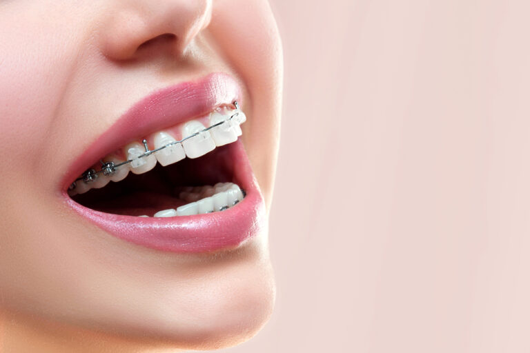
During orthodontic tooth movement, controlled forces loosen teeth within their sockets so they can shift positions. After treatment, stability typically returns as bone reforms. But this illustrates the potential mobility.
Age
As we age, our teeth often become slightly looser due to gingival recession exposing more mobile root surface and gradual diminishment of alveolar bone mass.
Signs that Tooth Mobility is Problematic

In most cases, the subtle movement of teeth when pressure is applied is harmless. But some key signs indicate that excessive mobility could stem from a pathological source:
- Tooth soreness, pain, or sensitivity
- Visibly noticeable or dramatic tooth movement
- Rapid progression of mobility
- Multiple loose teeth
- Known risk factors like gum disease or osteoporosis
If you exhibit any of these symptoms, it is best to promptly consult your dentist for an evaluation. Catching progressive mobility early is key to preserving tooth stability and oral health.
Protecting Tooth Support Structures
While normal mobility is expected, protecting the health of your gums, periodontal ligaments, and jawbone will help maintain that ideal balance of mobility and stability as you age. Preventive measures include:
- Practicing excellent oral hygiene with daily brushing, flossing, and professional cleanings to keep gums disease-free
- Using a nightguard if you are prone to clenching or grinding to minimize stress on teeth
- Getting orthodontic care to correct crowded, crooked teeth or problematic bites that overload certain teeth
- Limiting sugary foods and acidic drinks that damage enamel and contribute to decay
- Seeing your dentist regularly to identify any signs of increasing mobility early
With proper care, the complex and ingenious tooth support system will continue to allow for normal physiologic mobility without compromising the strength and integrity of your teeth.
Conclusion
Our teeth are designed for dynamic mobility as well as stability. Thanks to the periodontal ligaments and alveolar bone, teeth can withstand tremendous compressive forces during chewing while still exhibiting slight movement when other forces like tongue pressure are applied. This clever balancing act depends on healthy gums, bone density, and occlusal harmony to function optimally over a lifetime of use. A basic understanding of the anatomy and purpose of the tooth support structures can help explain why teeth wiggle but don’t fall out under normal conditions.
Expanded FAQ
Q: Are wiggly teeth always a sign of a problem?
A: No, slight wiggly movements when tongue pressure is applied are very normal and no cause for concern. True looseness involving visible shifting of teeth or continuous progression, however, can signal issues like periodontal disease. Context and degree matter.
Q: How much mobility is considered normal?
A: Normal physiologic mobility is up to 0.5 mm vertically and up to 0.2 mm horizontally. Anything beyond that range may indicate potential problems with bone or ligament support. One-directional jiggling movement is also normal, but multi-directional looseness is not.
Q: Do teeth ever fuse directly to bone without a periodontal ligament?
A: No, the PDL will always exist between the tooth root and jawbone. It serves an important function as a shock absorbing buffer. Total fusion of tooth to bone would make teeth more brittle and prone to fractures under oral forces.
Q: Can forceful tongue thrusting damage teeth over time?
A: Yes, if done repeatedly and excessively over many years, persistent forceful tongue thrusting or pressure can strain and micro-traumatize the front teeth. This risk is highest for upper incisors with smaller roots. Moderation is key.
Q: Why do front teeth seem to move more than back teeth?
A: Incisors and canines often have smaller root surface area and are shaped to have more of a mobile fulcrum effect. They trade stability for esthetics and specialized function in incising. Molars have broader roots and multiple anchorage points conferring greater stability.
Q: Is gum disease the only thing that causes abnormally loose teeth?
A: No, trauma like blows to the mouth can damage the PDL and periodontal support. Clenching and grinding over time also chronicly weakens the anchoring tissues. Some medical conditions like osteoporosis or drugs that cause bone loss can make teeth looser too.
Q: At what age do teeth typically start becoming looser?
A: Most people begin experiencing gradual age-related increased mobility in their 40s and 50s. Receding gums expose more mobile root surface, and cumulative oral loading combined with declining bone density both contribute. Keeping up with dental care helps slow this process.


