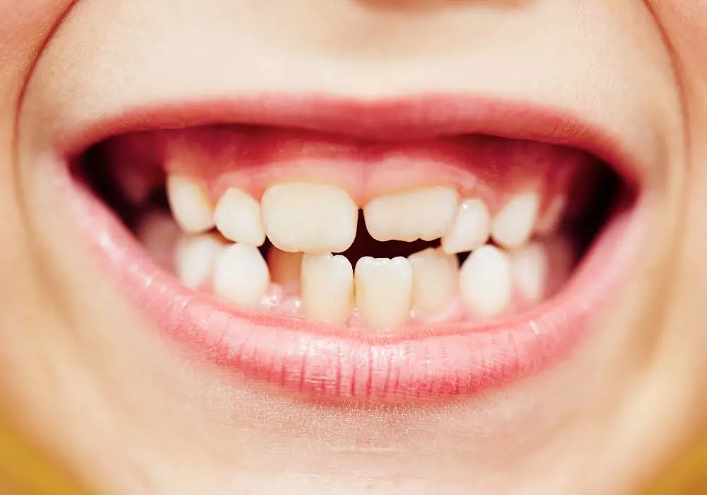
Noticing that your two upper front teeth are becoming progressively more transparent or translucent can be alarming. However, some degree of translucency in the incisors is normal as we age and enamel thins. More concerning are cases where the two central incisors exhibit accelerated transparency compared to other teeth, or have pronounced chalky spots or streaks.
There are numerous reasons this localized translucency may develop, including dental caries, erosion, fluorosis, trauma, tetracycline staining, developmental defects, and others. Determining the exact cause is crucial for proper treatment and prevention of further progression. Diagnosis requires a combination of visual inspection, dental imaging, patient history, vitality testing, and advanced assessment tools.
Based on etiology, treatment aims to address active disease, restore lost tooth structure, improve esthetics, prevent complications, and monitor teeth closely over time. While positive outcomes are possible with prompt intervention, certain types of enamel and dentin defects cannot be completely reversed.
Causes and Contributing Factors

Translucency isolated to the two maxillary central incisors has distinct characteristics and clinical findings that point to the underlying reason:
Dental Caries
Dental caries manifesting as barely noticeable frosty or opaque spots represents early demineralization of enamel minerals by oral bacteria. As acid dissolving progresses, white spot lesions develop which may transition into darker cavitated decay without intervention. Radiographs help detect areas of demineralization not visible clinically.
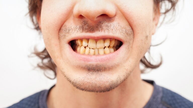
Cavitated caries confirmed as the cause reflects more advanced destruction into the enamel and dentin layers. This typically appears as yellow, brown or black softened areas of broken down tooth structure. Risk factors for caries like poor diet, dry mouth, poor oral hygiene, high acid foods and many carbohydrates are often present.
Chemical Erosion
Repeated exposure to acidic foods, beverages, stomach acids or environmental acids causes generalized thinning and surface defects rather than localized cavities. Underlying conditions like bulimia and gastric reflux increase risk, as do lifestyles with frequent acidic exposures. Enamel appears smooth, worn, and slightly transparent. Cupping, flattening or notching of the enamel surface may also be seen.
Enamel Hypoplasia
Systemic issues and medications like high fever, malnutrition, infectious diseases, trauma to developing tooth bud, chemotherapy, and anti-epileptics during infancy and early childhood disrupt enamel formation. Result is undermineralized enamel prone to defects like translucency. Medical history helps identify contributing factors.
Fluorosis
Overconsumption of fluoride while enamel is still developing causes subsurface porosities. This appears as barely noticeable fine white lines, streaks or flecking in mild forms, becoming more pronounced translucency and surface pitting in advanced cases. Fluorosis is confirmed through environmental exposure history, drinking water analysis, supplements, and observations of all teeth.
Trauma
Cracks, fractures, displacement injuries in early childhood can damage developing enamel. Chipping injuries later in life remove enamel thickness. Clenching and grinding worsens these post-traumatic defects over time. Clinical and radiographic exams locate cracks and assess pulp vitality. Prior trauma history is gathered.
Tetracycline Staining
Tetracycline incorporation into forming enamel and dentin when taken during tooth development causes yellowish to greyish translucency with brighter banding under UV light. The two central incisors are most affected due to calcification timing. Verifying tetracycline exposure from infancy to early childhood confirms diagnosis.
Amelogenesis Imperfecta
This genetic condition disrupts normal enamel thickness, mineral content, and structure. Teeth appear translucent, discolored, hypersensitive and prone to breakage. Defects are generalized affecting all teeth but most noticeable in upper anteriors. Family history and clinical characteristics are suggestive.
Dentinogenesis Imperfecta
Genetic defects in dentin formation result in teeth that are blue-grey or yellow brown in color with amber translucency concentrated in the incisal third and thinned enamel. They fracture easily due to brittle dentin. Positive family history and presence of osteogenesis imperfecta can aid diagnosis.
Internal Root Resorption
Damage to the pulp tissue inside the tooth triggers inflammatory destruction and resorption of root canal dentin first and then enamel from inside. Visible as a pinkish spot shining through enamel, this condition requires radiographs to confirm size, location, and progression over monitoring visits. Vitality testing detects nerve damage.
Diagnostic Workup
Because tooth translucency has many possible causes, accurate diagnosis requires a systematic approach:
- Comprehensive History – Medical conditions, dental history, fluoride exposure, tetracycline use, trauma, family genetics, enamel development issues, dietary factors and oral habits are documented.
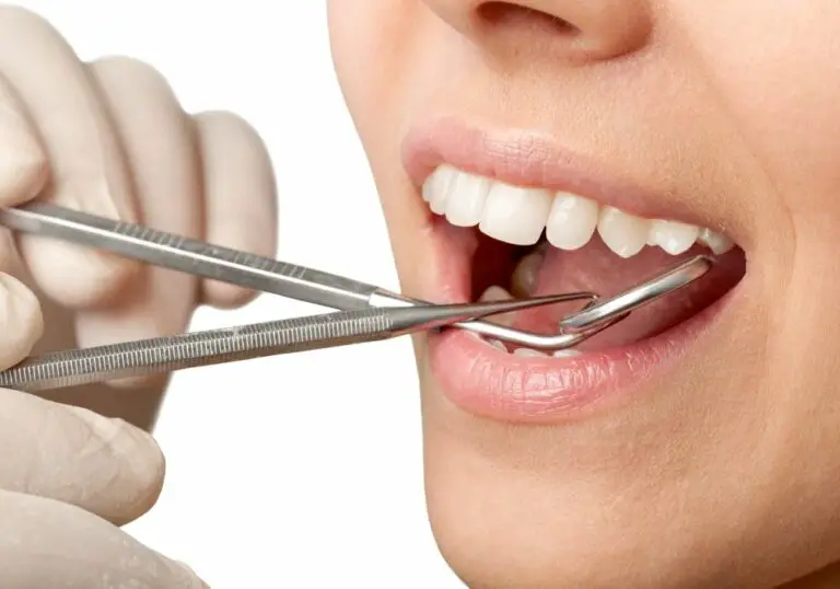
- Visual Inspection – Color, surface texture, location and pattern of translucency, presence of cracks, and light reflectivity are assessed clinically. Transillumination can aid detection. Photographs track changes over time.
- Dental Radiographs – Bite wing, periapical and if needed CBCT images identify subsurface decay, enamel defects, pulp health, mineral density, root and bone integrity, and check for other anomalies.
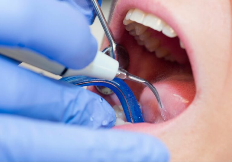
- Tactile Exam – Exploring tooth surfaces with a dental probe helps detect cracks, defects, cavitations, and surface roughness characteristics.
- Pulp Vitality Testing – Cold and electric pulp tests determine pulp health and nerve damage. Lack of response in just the two upper front teeth suggests possible resorptive process or trauma.
- Advanced Caries Detection – Tools like quantitative light induced fluorescence (QLF) and optical coherence tomography (OCT) objectively detect and monitor early demineralization over time.
- Acid Biopsies – Analysis of tooth surface pH levels indicates acid dissolution patterns from dietary acids, gastric reflux, bulimia, or environmental exposures.
- Salivary Testing – Analysis of salivary flow, pH, and buffering capacity identifies risk factors for accelerated dental caries.
Treatment Goals and Approaches
The primary treatment objectives are to address active disease, restore form and function, improve appearance, prevent further complications, and monitor progress closely over periodic recall visits.
- Control caries – Removal of decay and stabilization with fillings or other restorations. Prevention includes dietary changes, oral hygiene instruction, fluoride, sealants, saliva stimulants or modifying other caries risk factors.
- Manage erosion – Identify and manage acid source, whether gastric reflux, frequent dietary acids or environmental. Avoidance, buffering rinses, and acid-resistant restorations help strengthen teeth.
- Mask discoloration – Bleaching internally and externally, porcelain veneers, crowns and other esthetic methods mask translucency from fluorosis, imperfecta, tetracyclines, or hypoplasia.
- Increase tooth structure – Lost enamel and dentin is rebuilt with direct resin fillings, inlays, onlays, overlays, or full coverage crowns. For fractures, reattachment of fragment is attempted first.
- Extract severely compromised teeth – Non-restorable caries, resorption and other severe defects may necessitate extraction and implant placement if tooth cannot be saved.
- Take preventive measures – Dietary changes, oral hygiene instruction, fluoride, saliva substitutes, mouthguard use, and regular dental visits sustain treatment results.
- Provide protective restorations – Stabilization splints, mouthguards, veneers, and crowns reinforce teeth, prevent fracture of compromised structures and protect against future trauma.
- Monitor high risk teeth – Fluorosis, hypoplasia, imperfecta and other developmental conditions leave teeth permanently vulnerable, requiring close monitoring and preventive care.
- Perform needed endodontics – Root canal therapy stops internal resorption. Trauma, cracks, and carious exposure necessitate endodontic care to retain natural tooth structure.
Long-Term Outlook

With early intervention for diagnosed causes and diligent home care and followup, positive outcomes are possible. However, some types of enamel or dentin defects cannot be completely reversed, requiring lifelong monitoring and maintenance. Eventually reassessing major restorations may become necessary.
While treated or stabilized translucency may not worsen, some degree of future transparency in the front teeth is natural with aging. Ongoing prevention and regular dental visits sustain results long-term by catching any new issues early. But compromised teeth remain vulnerable to breakdown, fracture, wear and injury, requiring lasting vigilance.
FAQ
Q: Can whitening treatments help transparent spots in my front teeth?
A: External bleaching is unable to add opacity. It lightens natural teeth color only. Internal bleaching of discolored teeth due to resorption or endodontic treatment may improve translucency minimally but cannot restore enamel thickness or strength. Veneers, crowns or direct composite bonding are better options.
Q: Why do only my front top teeth have transparent areas?
A: The maxillary central incisors erupt at around 6-7 years of age as the last adult teeth to complete mineralization. This makes them most susceptible to developmental disturbances, trauma, mineral loss, and incorporation of systemic toxins during final maturation. Their visible positioning also makes defects more noticeable.
Q: Should I be concerned about transparent areas at the top edges of my front teeth?
A: Thinning along the incisal biting edges is often the first evidence of wear, acid erosion, or structural fatigue from malocclusions and bruxism. Further breakdown follows in the translucency zones, indicating a need for occlusal protective measures, acid reduction, and monitoring for fractures needing restoration.
Q: What is the best way to monitor transparent spots in my teeth?
A: Tracking with intraoral camera photographs, serial bitewing radiographs, and advanced diagnostics like quantitative light fluorescence, optical coherence tomography, and 3D CT scans quantitatively assess early demineralization before it becomes clinically visible. This allows preventive intervention before translucency progresses.
Q: Can dental bonding help strengthen my see-through front teeth?
A: Bonding repairs cracks, gaps, uneven edges, and small areas of breakdown but cannot increase structural durability like a crown. It is also prone to chipping and debonding under bite forces. Porcelain or ceramic restorations are stronger options for reinforcing severely compromised teeth.