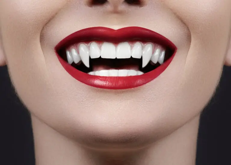
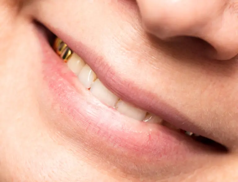
Canine teeth, also known as cuspids, eye teeth, or fangs, are the pointed teeth located on each side of the incisors in both the upper and lower jaws. Along with incisors and molars, canines are one of the three main types of teeth in mammals.
Canine teeth serve very important functions – they are used for piercing, tearing and holding food. Their sharpness and strategic placement in the dental arch gives them a crucial role in initiating the breakdown of food during mastication. Canines also help maintain proper alignment and occlusion with other teeth. But what exactly should normal, healthy canine teeth look like? In this detailed article, we will explore the anatomy, location, size, shape and appearance of canine teeth and what to watch out for.
Detailed anatomy of canine teeth
Canine teeth have a distinct anatomy optimized for their specialized role. Let’s look at the key structures in detail:
Root
The visible crown of a canine tooth is attached to the underlying jawbone by means of one or more tapered tooth roots. The root provides stability and support to the crown.
- Number of roots: Maxillary (upper jaw) canines usually have a single root. Mandibular (lower jaw) canines often have two roots placed close together.
- Length: Canine tooth roots are the longest among anterior teeth. The roots are deeply embedded in the alveolar bone to provide anchorage. Upper canine roots measure 16-22 mm on average. Lower canine roots are around 13-19 mm long.
- Shape: The root has a conical shape that is narrowest at the apex at the tip and progressively widens towards the cervix near the gum line. The surface is covered with an inert material called cementum.
- Internal canal(s): Running through the center of the root is the root canal, which houses soft tissue called the pulp and conveys the blood vessels and nerves. Mandibular canines often have two parallel root canals.
Enamel crown
The visible portion of the canine tooth that projects above the gum line is known as the anatomical crown. It is covered with enamel – the hardest substance in the human body.
- Total crown height: Maxillary canines have crowns 16-23 mm tall. Mandibular canine crown height is around 15-22 mm.
- Cusp height: The maxillary canine cusp projects around 10-12 mm vertically above the CEJ (cementoenamel junction). The mandibular canine cusp height is approximately 8-10 mm.
- Shape: Maxillary canines are shovel-shaped, while mandibular canines have a spearhead profile. The sharp cusp tip narrows incisally to blunter marginal ridges.
- Thickness: The labial enamel of canine crowns ranges from 1.5 – 2 mm in thickness. Palatal/lingual enamel is thinner at 1 – 1.5 mm.
- Enamel rods: Millions of closely packed enamel rods create the tough outer layer. The rods run obliquely from the dentinoenamel junction towards the tooth surface.
Dentin and pulp
Underneath the enamel layer is softer dentin surrounding the inner pulp chamber.
- Dentin thickness is around 1.5 – 2 mm along the crown. It appears darker than enamel due to lower mineral content.
- The pulp contains nerves and blood vessels entering through the root canals to nourish the tooth. It takes up around a quarter of the crown volume.
Periodontal apparatus
The root is attached to the bone via the periodontal ligament (PDL). The PDL fibers help anchor teeth firmly.
- Alveolar bone: The socket walls in the maxilla and mandible hold the tooth root in place.
- Gingiva: The gum tissue surrounds the cervical margin of the tooth crown.
- Cementum: This covers the dentin surface of roots and interfaces with the PDL.
Detailed location and placement of canine teeth
The pairs of canine teeth are strategically positioned in the upper and lower dental arches:
Maxillary canine location
- Situated lateral to the lateral incisors in the upper arch, with one canine on the left and right side.
- The maxillary canine crowns lie distal to the upper midline. The cusp tips are directly under the infraorbital foramen opening beneath the eyes.
- Roots are deeply embedded in the maxillary alveolar process, anchored between the lateral incisor and first premolar.
- Maxillary canines are the cornerstones of the upper arch, vital for reinforcing the bite.
Mandibular canine location
- Located next to the lower lateral incisors, again with one right and one left canine.
- Positioned closer to the anterior midline than the maxillary counterpart. Crowns lie just distal to the mandibular lateral incisor edge.
- The lower canine roots are deeply implanted in the mandible between incisors and premolars.
- Mandibular canines provide essential incisal guidance during excursive movements.
| Tooth type | Maxilla | Mandible |
|---|---|---|
| Central incisor | 1,2 | 1,2 |
| Lateral incisor | 3,4 | 3,4 |
| Canine | 5,6 | 5,6 |
| First premolar | 7,8 | 7,8 |
| Second premolar | 9,10 | 9,10 |
| First molar | 11,12 | 11,12 |
| Second molar | 13,14 | 13,14 |
| Third molar | 15,16 | 15,16 |
Detailed norm for canine size and shape
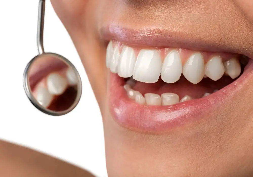
The canine teeth have a distinct size and form that matches their function:
Canine tooth length norms
- Maxillary canines are the longest teeth of the upper arch with crowns measuring around 25-30 mm visibly.
- Mandibular canines have the longest crowns in the lower arch, averaging approximately 20-25 mm in visible height.
- The upper canines are longer than the lower and protrude down beyond them by 2-3 mm when closed.
- Tooth length decreases progressively from canines to central incisors and premolars in both jaws.
Canine tooth width norms
- Maxillary canines are typically about 7-8 mm mesiodistally across the widest portion of their crowns.
- Mandibular canines average 5-7 mm in mesiodistal width.
- Upper canines are significantly broader than lower canines, which have a more blade-like form.
- The width of canines is greater faciolingually than mesiodistally, giving them their trademark robustness.
Typical canine shape and profile
The characteristic shovel vs. spearhead shape of maxillary versus mandibular canines is unmistakable.
- Maxillary canines have a strongly shovel-shaped crown whose palatal surface is markedly concave. The ridge is sharpened towards the cusp tip which comes to a blunted point.
- Mandibular canines have a slimmer, blade-like profile with a prominent vertical labial ridge and relatively flat lingual surface. The cusp tip is more sharply pointed.
- Viewed incisally, both arches have canines broader faciolingually, with rounded trapezoidal shapes.
Sexual dimorphism
Males tend to have more substantial and larger canine teeth than females, a form of sexual dimorphism. However, the overall shape remains similar.
Highly detailed canine tooth visible surface anatomy
The visible labial/buccal and incisal aspects of canine teeth provide much information about their health and status:
Labial/buccal surface details
- Enamel color – Healthy enamel is uniformly pale yellow to grayish white. Translucent bluish areas are normal near the cusp.
- Enamel thickness – Labial enamel is around 1.5-2 mm thick. Palatal enamel is thinner. Thickness affects translucency.
- Enamel texture – Surface is glossy smooth with fine perikymata (growth lines). No pitting, fractures or anomalies should be seen.
- Gum health – Pink, stippled gingiva should closely hug the cervical margin without swelling or recession.
- Root visibility – Cementum may show at the cervical area if gums have receded with age or periodontal disease.
Incisal edge details
- Cusp anatomy – Cusps should taper from incisal edge to marginal ridges smoothly without uneven wear facets or chips.
- Cusp tips – Maxillary cusps are more rounded versus sharper mandibular cusps. Cusp heights are 10-12 mm and 8-10 mm respectively.
- Incisal edge – Forms a clearly delineated continuous curve following the cusp contour. No dentin should be visible indicating wear.
- Incisal angles – The mesial and distal angles where the incisal edge meets the convexity of the tooth should be crisp, not rounded or worn.
Detailed canine tooth appearance changes from childhood to old age
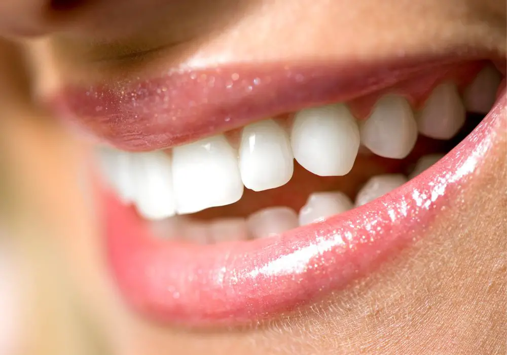
Canine teeth go through maturation changes from deciduous teeth to permanent replacements. Their appearance also evolves throughout life:
Canine teeth in childhood
- Deciduous canines – The upper and lower baby canine teeth are much smaller and more bulbous than permanent successors. Their thinner bluish enamel is distinctive.
- Permanent eruption – Maxillary permanent canines emerge around ages 11-13 years. Mandibular canines erupt earlier around 9-12 years. The existing deciduous canine is shed.
- Newly erupted – The fresh permanent canines are whiter and have cusp tips that become rapidly blunted from biting on eruption. Gingival contours are peaked around erupting teeth.
Teenage canine teeth
- By the late teens, maxillary and mandibular canines are fully erupted into place.
- Enamel is at its thickest and glossiest. The color is generally light yellow-white during this peak stage of maturity.
- Cusp tips become progressively worn to make a flatter incisal edge contact with lower canines.
- Canine prominence and high smile lines are often seen as gingival margins are at their highest position.
Adult canine teeth
- Early-mid adulthood sees canines relatively stable in the dental arch.Slow abrasion of cusp tips continues leading to gradual blunting.
- Chipping or cracking is possible with traumatic bites on hard foods. Fracture risk increases if bruxism wears down enamel.
- Discoloration can start with intake of chromogenic foods, smoking, medications, fluorosis or restorations.
Senior canine teeth
- Advancing age brings progressive thinning and increased transparency of enamel due to microscopic wear and tear.
- Dentin darkening leads to yellowing of the tooth crown and body. Pulp chamber shrinkage is common.
- Gingival recession exposes darker cementum near the cervical margin as ligaments weaken.
- Higher risk for cavities, abnormal wear, periodontal disease, and need for dental work like crowns, implants, bridges.
Detailed variations in normal canine appearance
Even when formed perfectly, no two canines are exactly identical. Some degree of minor variation can be considered normal between individuals and contralateral sides:
- Tooth dimensions – Subtle natural differences of a millimeter or two in visible crown length, cusp height or width.
- Crown contours – Smoothly shovel-shaped upper canines may appear bulkier than narrower spearhead lower canines.
- Cusp anatomy – Details of cusp ridge shape, tip rounding, marginal ridge shape vary.
- Enamel thickness – Thicker enamel has a more opaque white color vs thinner translucent enamel showing some underlying dentin shadow.
- Wear patterns – Asymmetric wear of cusp tip and incisal edge due to individual bite force distribution.
- Tooth alignment – Minor rotations or spacing may exist without constituting malocclusion.
- Restorations – Well matched veneers, crowns or implants mimic natural tooth form.
Highly detailed abnormalities and issues that can affect canine appearance
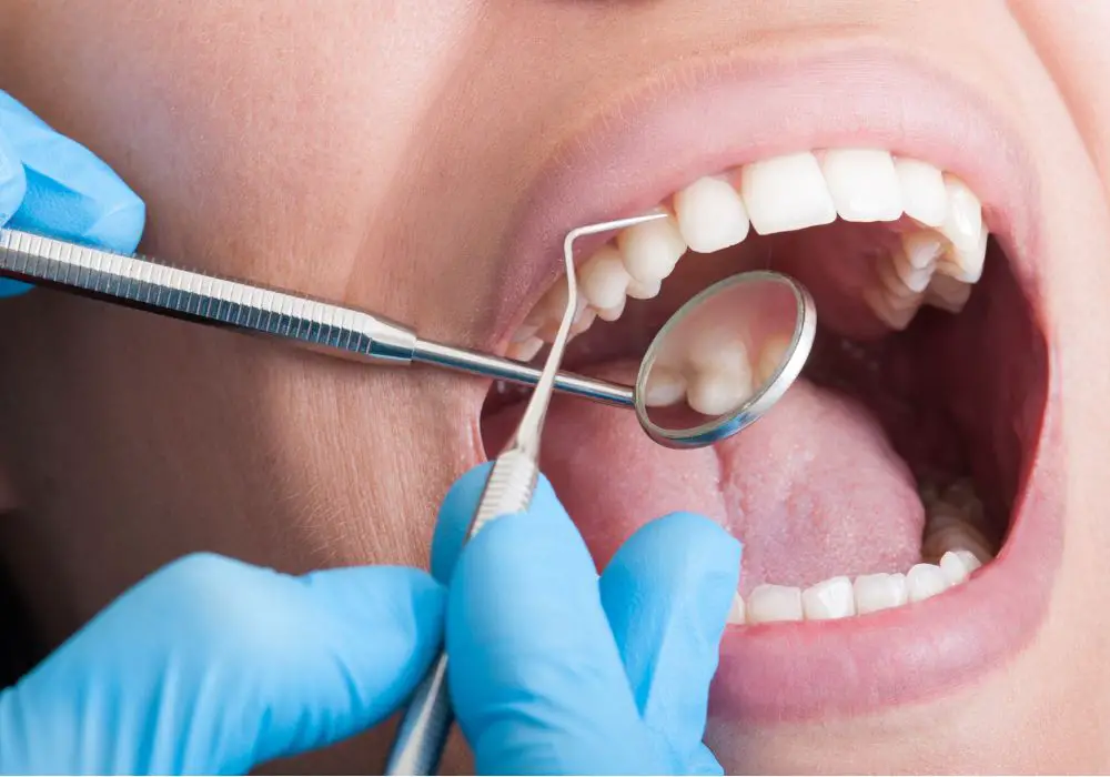
Canine teeth are vulnerable to a number of developmental or acquired issues that can alter their form and function from the norm:
Eruption problems
Failure of permanent canines to erupt properly is relatively common:
- Impaction – Lack of space or obstruction causes the unerupted canine to be stuck in bone. This also prevents shedding of the persistent deciduous canine.
- Retention – The deciduous canine remains beyond age 14-15 years due to failure of the permanent successor to erupt into place.
- Ectopic eruption – The permanent canine emerges in the wrong orientation or position due to lack of space or developmental anomalies.
- Transposition – Upper canine switches position with first premolar or lateral incisor during development.
Injuries and excessive wear
Dental trauma or parafunctional habits can damage canine teeth:
- Crown and root fractures – Cracks, chips or complete fractures due to blows or accidents. May require crowns or extraction.
- Attrition – Wearing away of enamel and dentin from grinding and clenching, especially on the cusp tips and incisal edge.
- Abrasion – Loss of tooth structure from mechanical abrasion as from overzealous tooth brushing.
- Erosion – Chemical dissolution of enamel from dietary acids in beverages, citrus fruit etc.
Discoloration issues
Discoloration of canine teeth can have varied causes, including:
- Dental fluorosis – Overexposure to fluoride during crown formation leads to white opaque streaks or mottling.
- Tetracycline staining – Systemic tetracycline antibiotic intake during tooth development causes yellow to dark grey discoloration in bands.
- Pulp necrosis – Death of the pulp vessels turns the crown darker. Typically requires root canal therapy or extraction.
- Dental caries – Bacterial decay causes cavitations and brown discoloration penetrating the enamel.
Developmental anomalies
Genetic conditions can alter canine morphology:
- Amelogenesis imperfecta – Defective enamel formation leads to thin, weak, discolored enamel prone to defects.
- Dentinogenesis imperfecta – Mutations affect dentin formation, causing translucent, structurally weak teeth.
- Supernumerary canines – Extra canine teeth, either a supplemental tooth or bilateral pairs.
- Dens invaginatus – Deep enamel folding within the crown during development, making it prone to decay.
- Dilaceration – Trauma to the forming permanent tooth causes deviation of the developing root apex.
- Gemination- An attempt to divide into two conjoined crown structures during development.
Professional evaluation is critical for abnormal canine teeth
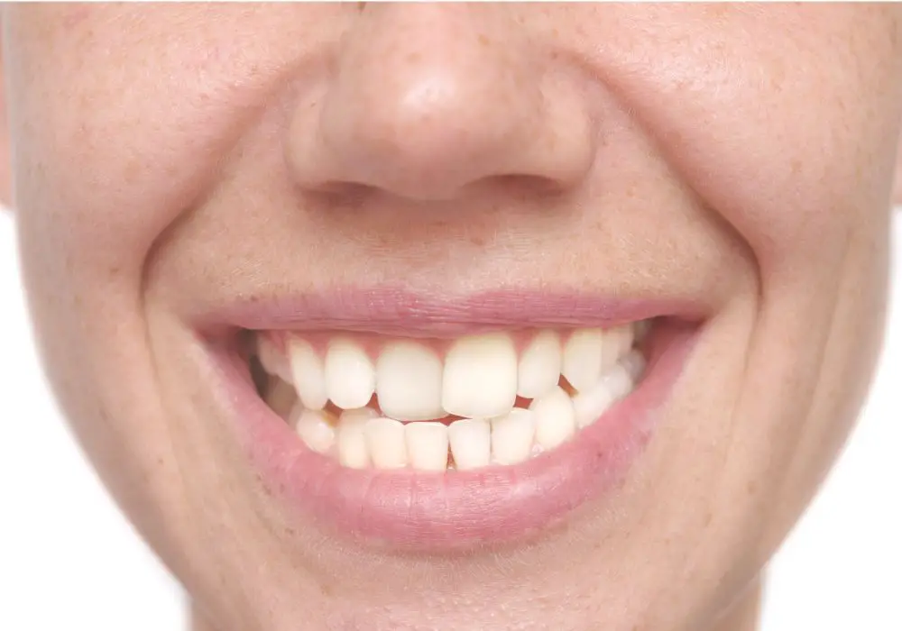
Changes in canine appearance, especially discoloration, chipping, wear or shape irregularities should prompt evaluation by your dentist. They can:
- Diagnose the underlying issue based on clinical and radiographic findings.
- Determine whether restorative procedures, root canal therapy, extractions or other treatment is required.
- Provide preventive guidance to avoid further damage and preserve remaining tooth structure.
- Recommend cosmetic correction with crowns, veneers or implants for greatly compromised teeth to restore form and aesthetics.
Routine dental exams every 6 months also allows early detection and monitoring of problems. Seek prompt help for any trauma, pain or visible fracture involving canine teeth. Addressing issues early improves outcomes.
Extensive preventive measures to maintain canine health
Preserving natural, healthy canine teeth function requires diligent oral hygiene and smart lifestyle habits:
- Brush carefully – Use a soft-bristled toothbrush and fluoride toothpaste. Take extra time on canine surfaces. Angle brush to access incisal edge.
- Floss thoroughly – Use waxed floss or threaders to wrap around and hug each canine tightly to dislodge plaque.
- Professional cleanings – Tartar removal and fluoride application protects canines from decay. Alert your hygienist to any sensitivity.
- Balanced diet – Avoid excessive acidic and sugary foods that erode enamel or promote cavities.
- Chew wisely – Don’t bite or tear with your canines. Cut hard foods into pieces first.
- Wear mouthguards – Protect canines from sports injuries or bruxism damage. Nightguards shield from nocturnal grinding.
- See your dentist – Get regular oral cancer screenings. Report any chipping, pain, swelling or color changes for evaluation.
Answers to frequently asked questions about canine teeth
Here are some common questions and detailed answers about canine teeth:
Why are canine teeth so pointy?
The sharp, prominent cusps of canine teeth serve to pierce into food. Their narrow tips concentrate force for biting and tearing foods efficiently. The pointed shape also enables canines to grip and hold objects firmly.
What problems may occur if canines are lost or damaged?
Canines help guide the bite and stabilize the dentition. Loss of canines can shift other teeth out of alignment. It can also reduce chewing efficiency and power. Damaged canines are more prone to wear, fracture and pulp issues.
How does canine function differ between upper and lower arches?
Maxillary canines are adapted for piercing and holding foods in place as the lower canines shear more forcefully. Their roots absorb pressure during biting. Mandibular canines provide a vertical stop

