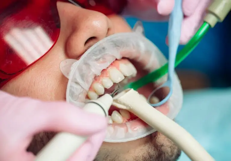
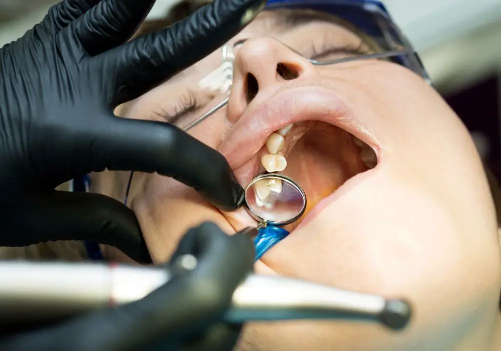
The upper jaw, also known as the maxilla, is one of two main bones that make up the upper part of the mouth and face. It contains the upper teeth and helps form the palate, nasal cavity, and sinus cavities. Understanding the anatomy and function of the upper jaw is key for dental health. This comprehensive guide will examine in detail if and how the upper jaw contains teeth.
Detailed Anatomy of the Upper Jaw
The upper jaw consists of two maxilla bones that fuse together during embryonic facial development. Here is a more in-depth look at the anatomy:
- Each maxilla bone has a body, frontal process, zygomatic process, palatine process, and alveolar process.
- The body forms the anterior lateral nasal wall, inferior orbital wall, and the lateral palate.
- The frontal process articulates with the nasal bone and lacrimal bone.
- The zygomatic process articulates posteriorly with the zygomatic bone.
- The palatine process meets medially to form the anterior hard palate.
- The alveolar processes contain the upper tooth sockets.
- The maxillary sinus is a cavity within each maxilla body. It pneumatizes over time.
- Specialized cells produce the bony matrix while osteoclasts resorb bone during growth.
- Many muscles and ligaments anchor to the upper jaw, enabling movement.
- Nerves and blood vessels supply the maxilla through infraorbital canals and foramina.
In summary, the complex upper jaw structure supports its functional role in housing the maxillary teeth and forming vital facial regions.
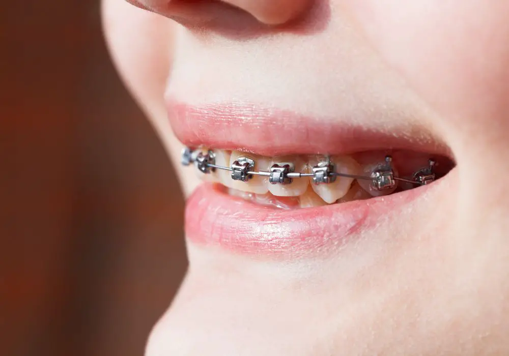
Upper Jaw Dentition Includes Both Primary and Permanent Teeth
The upper jaw holds two sets of teeth over a person’s lifetime:
- The primary (baby) teeth erupt first and include incisors, canines, and molars.
- These are replaced by the permanent teeth which also include premolars.
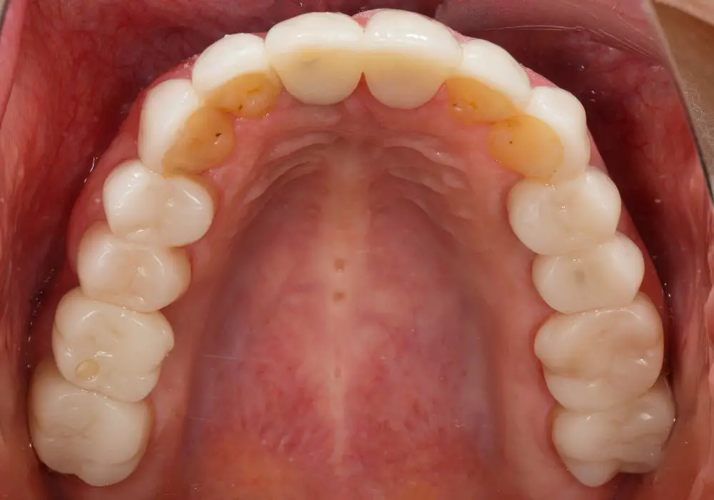
The 16 permanent maxillary teeth include:
- 4 incisors (2 central, 2 lateral)
- 2 canines
- 4 premolars (2 first, 2 second)
- 6 molars (2 first, 2 second, 2 third)
The primary upper teeth are smaller with thinner enamel and root structure adapted for shedding.
The permanent teeth are larger, with thicker dentin and enamel. They utilize longer, multi-rooted anchorage.
Both primary and permanent dentitions allow for functional chewing, speech, and tooth-to-jaw proprioception.
Alveolar Bone Anchors Teeth to the Upper Jaw

The upper jaw supports each tooth through specialized alveolar bone:
- The lamina dura lines the tooth socket (alveolus) next to the root.
- It is covered by a thin layer called the bundle bone.
- The spongy bone of the alveolar process surrounds this.
- The periodontal ligament fibers embed within the bone matrix.
- Alveolar bone has a high turnover rate and remodeling capacity.
- Osteoblasts and osteoclasts balance bone deposition and resorption.
- Proper alveolar bone volume maintains the teeth in good alignment.
- Loss of alveolar bone can lead to loosening or loss of teeth.
Thus, the structured alveolar bone of the maxilla firmly yet flexibly anchors each tooth.
Unique Growth Pattern for Upper Jaw and Teeth
Three dimensional growth of the upper jaw allows for the coordinated emergence of teeth:
- The maxilla descends and remodels most significantly during childhood.
- Growth hormone and genetic factors control sutural growth at joints.
- Muscles like the masseter provide strain to stimulate bone deposition.
- The palatal processes fuse by the bud stage of first molar formation.
- Teeth sequentially erupt through programmed formation and eruption patterns.
- First molars erupt around age 6, anchoring posterior occlusion.
- Eruption of incisors and canines follows, establishing anterior guidance.
- The jaw grows outward as later teeth develop more posteriorly.
This intrinsic maxillary growth results in a balanced, functional upper dental arch.
Impact of Congenital Clefting on Upper Jaw Dentition
Clefts of the upper jaw and palate are common congenital disorders that affect tooth development:
- Clefts occur where maxillary processes fail to fuse in the embryo.
- Cleft palate disrupts the palatine shelves uniting.
- Alveolar clefts directly split the upper dental arch.
- Tooth buds can be missing, malformed, displaced or impacted.
- Supernumerary teeth are also common with clefting.
- Eruption sequence is delayed and dysfunctional.
- Malocclusion and open bite often result due to imbalance.
- Special orthodontic appliances and surgical repair are needed.
In summary, clefting can significantly impair proper dentition within the upper jaw.
Conclusion
In conclusion, the upper jaw certainly does contain the maxillary teeth, with both primary and permanent dentitions held by specialized alveolar bone. The complex growth and maturation of the maxilla allows for the coordinated emergence of correctly aligned, functional upper teeth. However, congenital disorders like clefting can disrupt normal upper jaw dentition and must be treated early with reconstructive methods. Overall, the anatomy and development of the upper jaw provides the foundation for housing the upper teeth.
Expanded FAQ
Q: How many teeth are in the upper jaw?
A: There are 16 permanent teeth in the upper jaw – 2 incisors, 2 lateral incisors, 2 canines, 4 premolars, and 6 molars. The primary upper jaw has only 10 teeth.
Q: What bone contains the upper teeth?
A: The maxilla, or upper jawbone, contains the upper teeth within its alveolar processes and alveolar bone sockets.
Q: Do the upper front teeth have a special name?
A: Yes, the four upper front teeth (central incisors and lateral incisors) are also known as the maxillary anterior teeth or maxillary incisors.
Q: Can a cleft palate affect the upper teeth?
A. Yes, a cleft palate can displace, deform, or impact upper teeth since it splits the upper jaw structure. This can lead to missing, irregular, or malaligned teeth if not corrected.
Q: How does the upper jaw change with age?
A: The upper jaw grows most significantly in childhood and becomes stable in adulthood. The alveolar bone begins to resorb after tooth loss, reducing jaw height. Density decreases after age 40. Sinus cavities expand.
Q: Can dental implants be placed in the upper jaw?
A: Yes, the upper jaw provides excellent stability for dental implants if alveolar bone density is sufficient. Implants fuse to the maxillary bone to replace missing teeth.
Q: What is the recommended age for upper jaw growth completion?
A: Maxillary growth is largely complete by age 18 in females and age 20 in males. Minor changes can occur through the mid-20s but this marks general maturation of the upper jaw structure.
Q: Does the maxillary sinus affect the upper back teeth and jaw?
A: Yes, the maxillary sinus expands via resorption, particularly after upper molar extractions. This can reduce alveolar bone height and lead to pneumatization of the sockets.