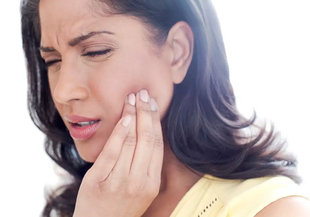
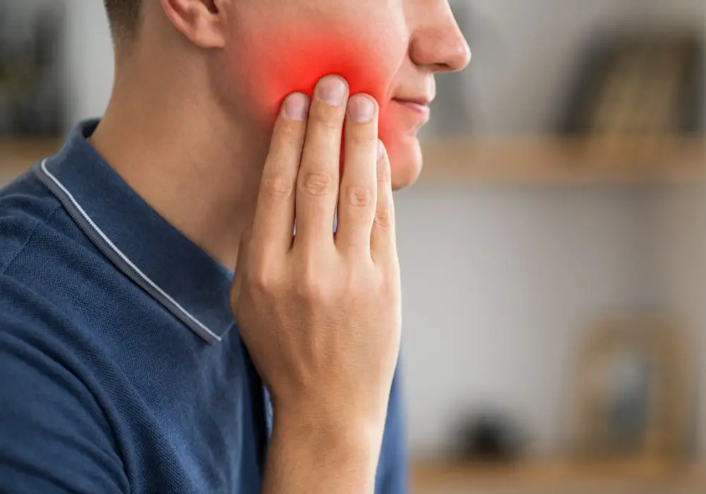
Oral health is determined by a complex combination of genetic, epigenetic, environmental, and behavioral factors. While diet, trauma, smoking, and access to care all impact dental disease risk, there are also a number of conditions directly caused by inherited genetic mutations or traits passed down in families. This article will provide an in-depth overview of the main hereditary dental diseases and developmental disorders affecting tooth structure, enamel and dentin formation, tooth eruption and alignment, and susceptibility to dental caries and periodontal disease.
What Dental Diseases are Hereditary?
Enamel formation defects
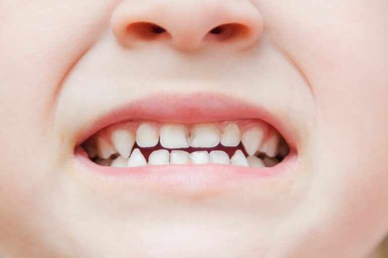
Tooth enamel is the remarkably hard, crystalline mineral outer layer that covers and protects the anatomy of each tooth crown. Enamel is unique among tissues in that it does not remodel or regenerate itself once formed, so any defects during initial development permanently alter its structure and strength. Enamel formation occurs in stages under tight genetic control, with ameloblasts cells secreting specific matrix proteins that direct the mineralization process and maturation of hydroxyapatite crystals.
Amelogenesis Imperfecta
Amelogenesis imperfecta represents a group of hereditary conditions causing defective tooth enamel formation, resulting in enamel that is thin, soft, rough, and prone to rapid decay and breakdown. It is caused by mutations in the genes coding for enamel matrix proteins like amelogenin, enamelin, enamelysin, and ameloblastin which are essential for proper enamel development. The defective enamel cannot withstand the normal wear and acid dissolution from oral bacteria that occur in the mouth. There are four main phenotypic presentations of amelogenesis imperfecta:
- Hypoplastic – Thin, hard enamel lacking sufficient thickness and volume. Caused by mutations in enamel-specific genes like AMELX, ENAM, KLK4. Autosomal dominant inheritance.
- Hypomaturation – Defectively mineralized enamel that is softer, with lower hydroxyapatite content. MMP20 and KLK4 gene variants often implicated. Autosomal recessive inheritance.
- Hypocalcified – Normal enamel thickness but very low mineral content leaving it soft and easily removed. Mutations affecting FAM83H. Autosomal dominant inheritance.
- Hypomature-hypoplastic – Enamel is thin with deficient mineralization. Caused by WDR72 mutations. Autosomal recessive inheritance.
Over 10 different gene loci have now been identified that can cause non-syndromic amelogenesis imperfecta inheritance when enamel defects occur in isolation without other syndromic symptoms. X-linked, autosomal dominant, and autosomal recessive inheritance patterns are seen, depending on the specific genes involved. Genetic testing and pedigree analysis allows determination of the precise causative mutations within families affected by this enamel defect disorder.
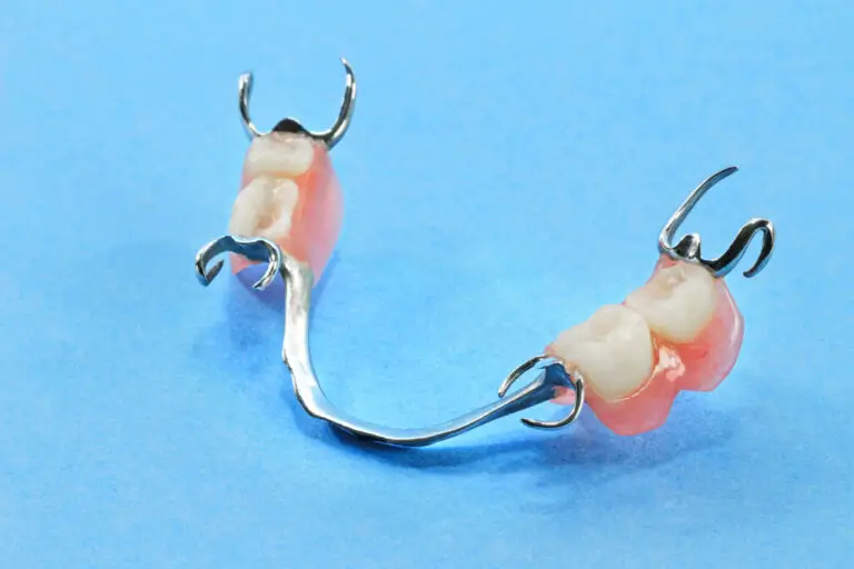
Treatment for amelogenesis imperfecta is aimed at protecting the weakened, sensitive enamel from additional breakdown. Excellent oral hygiene, fluoride exposure, dietary modifications, and resin restorations for badly damaged teeth help prevent rapid tooth destruction and loss. However the defective enamel remains permanently vulnerable despite intervention efforts.
Dentinogenesis Imperfecta
Analogous to amelogenesis imperfecta, dentinogenesis imperfecta is an inherited disorder causing defective dentin formation, resulting in teeth with amber-brown or blue-gray discoloration that are prone to wear, fracture, and pulpal problems. Abnormal dentin structure results from mutations affecting the collagen matrix proteins secreted by odontoblast cells during dentin development under the enamel layer.
Type 1 dentinogenesis imperfecta with amber-brown teeth is caused by autosomal dominant mutations in the DSPP gene encoding dentin sialophosphoprotein. Type 2 dentinogenesis imperfecta with blue-gray teeth has autosomal recessive inheritance often linked to DSPP recessive mutations, but can also involve DDPII and COL1A1 collagen gene defects. Severe cases can present with osteogenesis imperfecta systemic connective tissue defects.
As with enamel defects, the aberrant dentin morphology and mineral content caused by these genetic mutations lead to rapidly deteriorating dental health even with diligent oral hygiene efforts. Management involves diligent fluoride treatments and dental repair or restoration of fractures, but the dentin defect persists.
Developmental dental disorders
Ectodermal Dysplasia
Ectodermal dysplasia represents a group of over 150 distinct congenital syndromes caused by genetic defects affecting ectodermal derived tissues like skin, hair, nails, sweat glands, and crucially for dental health – teeth. Mutations in signaling factors like EDA, EDAR, and WNT10A disrupt reciprocal epithelial-mesenchymal interactions during tooth development, leading to missing teeth (hypodontia), malformed pointed or cone shaped teeth, and lack of normal tooth enamel.
Most cases have X-linked recessive inheritance, but autosomal dominant and recessive inheritance also occur. In addition to dental defects, those affected have problems with sparse hair, reduced sweating, and skin pigmentation. Management of the oral manifestations aims to restore function and aesthetics via dental implants, adhesive restorations, and prosthetics.
Regional Odontodysplasia
Also known as ghost teeth, this rare non-hereditary condition causes segmental enamel and dentin defects localized to just a few teeth, often in the same quadrant or region of the mouth. Affected teeth have a yellow-brown discoloration and are typically soft, porous, hypoplastic, and prone to rapid wear and decay. The cause is unknown but may involve vascular disruption or local trauma during tooth calcification in utero. Extensive restorations are often needed to salvage ghost teeth and prevent deterioration.
Dentin Dysplasia
Dentin dysplasia involves abnormal formation of root and crown dentin leading to short, blunted tooth roots and pulpal obliteration.Inherited dentin defects result in amber-brown or bluish-gray discolored teeth with a high risk of pulp necrosis. Root malformation also occurs. Autosomal dominant and recessive subtypes exist, caused by mutations in the DSPP gene on chromosome 4q. Tooth extractions are often unavoidably needed due to pulpal abscess and fracture risk.
Cleft lip and palate
Oral clefts including cleft lip, cleft palate, or both together have a strong hereditary component, with several identified genetic risk loci. Clefts occur when parts of the lip and palate fail to fuse together during embryonic craniofacial development due to disrupted migration of neural crest cells into the branchial arches that form the face and jaw structures.
This leaves openings that can involve the upper lip, the roof of the mouth (hard palate), and/or the soft tissue rear portion of the palate. Clefts often cause subsequent dental problems like missing teeth near the cleft site, malformed teeth, tooth eruption anomalies, and misalignment due to the altered bone and soft tissue anatomy.
Having a cleft increases risk for dental caries and periodontal disease due to the specialized orthodontic care and oral hygiene required. Surgical repair of the lip and palate, often accompanied by later bone grafting, can restore normal aesthetics and function but does not fully prevent dental complications like speech problems and misalignments.
Periodontal disease susceptibility
Aggressive Periodontitis
This severe and rapid form of gum disease causes rapid deterioration of periodontal ligament and alveolar bone in otherwise healthy individuals, typically striking during puberty or early adulthood. A very strong genetic component is implicated, with inheritance patterns often consistent with autosomal dominant transmission, although polygenic risks also likely contribute. Even with minimal dental plaque and excellent oral hygiene, aggressive periodontitis often leads to premature tooth loss without early treatment.
Multiple gene mutations affecting immune factors, inflammation regulation, and tissue destruction pathways appear to be related to increased susceptibility to aggressive periodontitis. Neutrophil defects may be involved, with abnormalities in HMPO, cathepsin C, and other lysosomal enzymes seen. Still, the specific genetic drivers remain unclear. However aggressive periodontitis does cluster strongly in families, confirming a powerful genetic influence separate from routine plaque induced chronic adult gum disease.
Chronic Periodontitis
The typical adult form of gum disease that develops slowly from persistent gingivitis also has genetic risk factors, but heavily influenced by modifiable environmental exposures like smoking and poorly controlled diabetes. Still, twin studies suggest approximately 50% heritability even for chronic periodontal disease, confirming an inherited susceptibility component. There seem to be genetic tendencies accounting for variation between individuals in immune response to oral pathogens, inflammatory regulation, and tissue destruction upon bacterial biofilm challenge.
Multiple subtle genetic variations involving cytokine levels (IL-1, IL-6, TNF-alpha), cellular receptors (toll-like receptors), and enzymes like collagenase have been associated with chronic periodontal disease risk and severity. Gene variants in the vitamin D receptor also appear implicated. However, diligent oral hygiene and managing other risk factors allows chronic gum disease to be prevented or minimized for most people regardless of genetics.
Dental caries susceptibility
Tooth decay is well established to result from a complex interplay between acid-producing oral bacteria like streptococci and lactobacilli, fermentable dietary carbohydrates, saliva properties, and the integrity of the tooth enamel surface. However, there is also evidence suggesting a genetic influence on dental caries risk. Some individuals seem prone to cavities despite careful oral hygiene, while others develop little decay even with poor brushing habits, implying stronger genetic resistance.
First degree biological relatives often share similar dental caries rates and patterns, pointing to a heritable susceptibility component. Specific genetic variations have been identified that affect enamel formation, saliva quantity and composition, tooth pit and fissure morphology, and the oral microbiome – all of which can modulate dental decay risk. Caries risk assessment incorporating genetic markers may someday help better predict decay susceptibility versus resilience to aid prevention approaches. For now, those with high heritable decay risk benefit from additional preventive therapies and vigilance.
Tooth agenesis and missing teeth
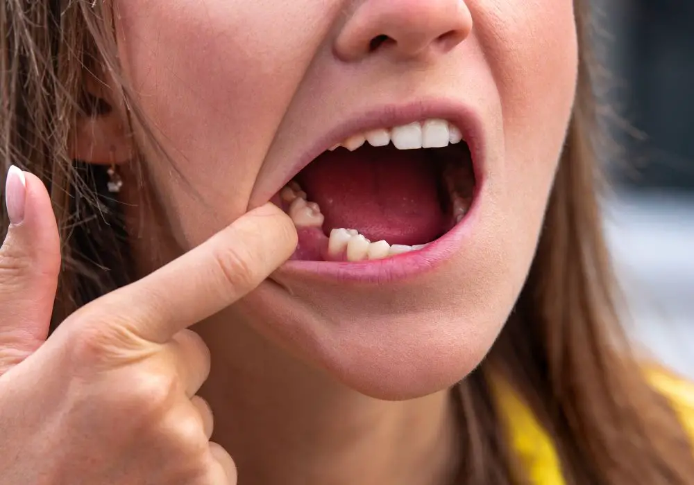
Non-Syndromic Hypodontia
This condition is defined as having 6 or more permanent teeth congenitally missing or unerupted, excluding third molars. The most commonly missing teeth are mandibular second premolars, maxillary lateral incisors, and maxillary second premolars. Hypodontia occurs in 3-5% of the general population to varying degrees, and is classified as mild (2-5 teeth missing), moderate (6-9 missing) and severe (over 9 missing).
Both environmental insults and genetic factors are believed responsible for hypodontia, with mutations in various genes impairing tooth follicle development involved. These include MSX1, PAX9, AXIN2, EDA, and WNT10A among others. The phenotypic expression and inheritance patterns of non-syndromic hypodontia are highly variable, consistent with polygenic risks interacting with epigenetic and environmental influences on tooth formation.
Oligodontia
Oligodontia represents a more severe form of developmental tooth agenesis characterized by 6 or more congenitally missing teeth, often including first and second permanent molars or other teeth that normally demonstrate very low rates of agenesis. This rare condition affects 0.1-0.2% of the population. The pattern of missing teeth can include incisors, canines, premolars, and molars.
Known genetic causes of isolated oligodontia typically involve mutations in MSX1, PAX9, and AXIN2, which are commonly implicated in isolated tooth agenesis. These appear to disrupt early tooth development, impairing bud initiation and subsequent mineralization stages. Oligodontia often shows autosomal dominant inheritance patterns within families, but other genetic loci may also be involved. This severe level of missing teeth requires extensive dental restoration and rehabilitation to establish functional occlusion.
Inherited oral disease syndromes

A number of genetic syndrome conditions exist that involve multiple symptoms affecting the whole body, including characteristic oral manifestations. These hereditary syndromes often involve dental developmental defects, clefting, and increased dental disease risks:
- Down syndrome – delayed tooth eruption, smaller jaws, periodontal disease, and unique mouth breather facial appearance
- Marfan syndrome – increased oral ulcers, root resorption, pulp calcifications, and hypermobile TMJ issues
- Ehlers Danlos syndrome – early onset periodontitis affects over 50% by age 20 due to tissue laxity and impaired wound healing
- Papillon Lefevre syndrome – severe early onset periodontitis with complete tooth loss by teens, linked to cathepsin C mutations
- Cleidocranial dysplasia – missing teeth, retained supernumerary teeth, and lack of complete jaw development
Key Takeaways
- Enamel defects like amelogenesis imperfecta have strong inherited genetic origins from mutations disrupting matrix protein secretion during enamel development.
- Dentin defects including dentin dysplasia and dentinogenesis imperfecta also arise from genetic errors affecting collagen matrix and mineralization.
- Developmental syndromes such as ectodermal dysplasia show dental manifestations of conical teeth, enamel defects, and severe hypodontia due to genetic mutations impairing reciprocal epithelial-mesenchymal interactions.
- Many other genetic syndromes have oral effects including altered dental development, increased caries and periodontal risks, and clefting.
- Caries and gum disease have complex etiologies involving interplay between genetic predisposition and modifiable exposures like diet and smoking.
- Aggressive periodontitis and conditions like Papillon Lefevre syndrome demonstrate very penetrant genetic origins, while chronic periodontitis and caries have weaker genetic links.
Frequently Asked Questions
Are cavities and tooth decay hereditary?
Tooth decay results from the interplay between oral bacteria, fermentable carbohydrates, saliva factors, and tooth enamel integrity. No single “cavity gene” exists. However, specific genetic variations affecting enamel, saliva composition, tooth morphology, and the oral microbiome likely modulate individual susceptibility versus resistance to dental caries. So genetic risks contribute but are not wholly deterministic for decay.
Is gum disease genetic? Are periodontal problems inherited?
Periodontal diseases have both genetic and environmental risk factors. Aggressive early-onset periodontitis has very strong genetic influence with predominantly autosomal dominant inheritance patterns in families. Chronic adult periodontitis is more complex with only around 50% heritability, meaning behavioral and health factors like smoking have large impacts. Still, variations in inflammatory genes and tissue enzymes do affect genetic susceptibility.
Why do malocclusions and crooked teeth run in families?
Tooth size, jaw dimensions, and growth patterns are all inherited and contribute to misalignments and dental crowding. Having a small jaw, large teeth, or overbite/overjet as in other family members makes crowded teeth more likely. However, oral habits, injuries, diet, and other factors also affect alignment. While genetics play a key role, orthodontics and surgery can help straighten teeth.
What is the genetic basis behind gum disease?
Research suggests certain gene variants affecting inflammatory mediators (IL-1, IL-6, TNF-alpha), matrix metalloproteinases like collagenase, immune cell receptors, and osteoclast activation likely underlie increased susceptibility to periodontal infections and tissue destruction. Mutations directly affecting neutrophil and monocyte function also contribute to severe early onset forms. Good oral hygiene remains essential to control genetic gum disease risks.
Why do problems like weak enamel seem to run in families?
Enamel formation is tightly genetically controlled by secreted proteins like amelogenin and enamelin. Mutations in the genes encoding these proteins lead to defective enamel formation and inherited conditions like amelogenesis imperfecta. Such enamel defects are passed down in families in X-linked, autosomal dominant or recessive patterns depending on the causative gene. Other issues like deeper fissures also have genetic links.
Conclusion
In summary, genetics play a very significant role in many different dental diseases and anomalies, particularly enamel and dentin developmental defects which arise directly from inherited mutations. Environmental influences like diet, smoking, and access to care strongly impact susceptibility to complex multifactorial conditions like caries and chronic periodontitis. However, genetic variations contribute to risk as well. Understanding the hereditary basis of certain dental problems allows better prediction of family risks and patterns, earlier diagnosis when conditions are familiar, and more customized prevention approaches. Still, daily oral hygiene remains essential even in the presence of genetic risk factors to maintain the best possible oral and systemic health.